Latin: Atrium sinistrum
Overview
- The chamber that receives oxygenated blood from the lungs through the four pulmonary veins.
- Forms most of the base (posterior surface) of the heart; passes blood to the left ventricle through the mitral valve.
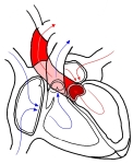
Chambers of the heart and the flow of blood
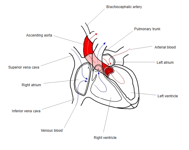
Features
- Mostly smooth-walled; pectinate muscles are found only in the left auricle.
- The septal wall shows the valve of the foramen ovale (the left side of the fossa ovalis).
Relations
- Lies directly in front of the oesophagus, separated from it by the pericardium (the oblique sinus).
Clinical notes
- An enlarged left atrium (for example in mitral stenosis) can press on the oesophagus.
- In atrial fibrillation, clots tend to form in the left auricle.
- Transoesophageal echocardiography views the left atrium closely because of this relation.
References and sources
- Further reading: Left atrium, Wikipedia.