Latin: Vena centralis hepatis
Overview
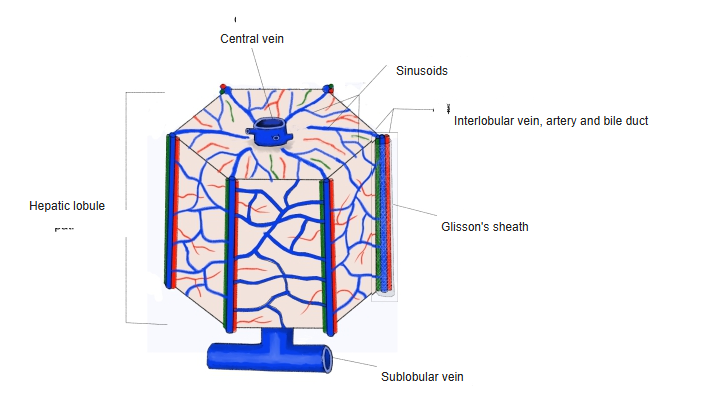
- A small vein in the center of each hepatic lobule.
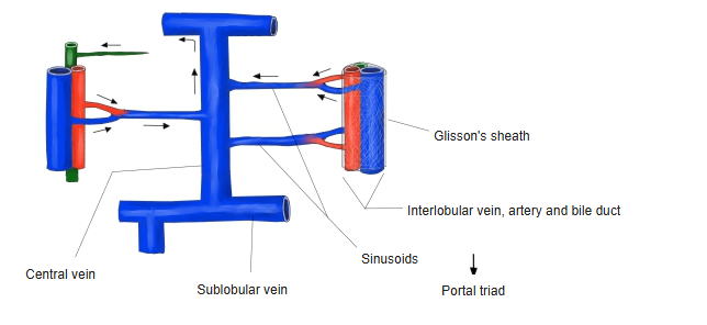
- Receives blood from the sinusoids, which carry mixed portal venous and hepatic arterial blood from the portal triads at the corners of the lobule.
- Central veins join to form sublobular veins, which unite into the hepatic veins.
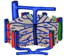
Blood vessels of the hepatic lobule 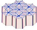
Hepatic lobule and central vein
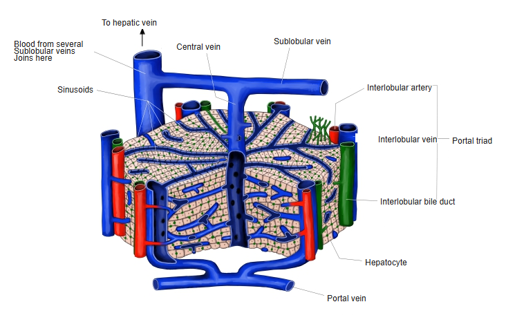
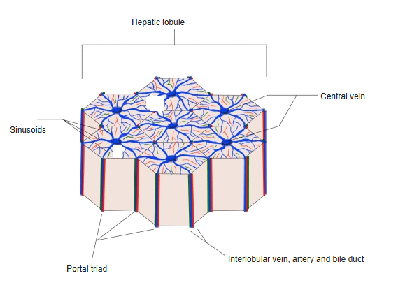
Hepatic lobule
- Plates of hepatocytes radiate from the central vein; the portal triads (interlobular vein, interlobular artery, bile duct) lie at the corners.
- Blood flows toward the central vein; bile flows the opposite way, toward the portal triads.
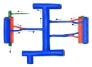
Hepatic lobule (model) 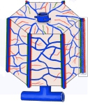
Hepatic lobule and sinusoids
Clinical notes
- Cells around the central vein (zone 3) receive the least oxygen and are the first damaged in heart failure and some drug injuries.
References and sources
- Further reading: Central vein, Wikipedia.