Latin: Vena portae hepatis
Overview
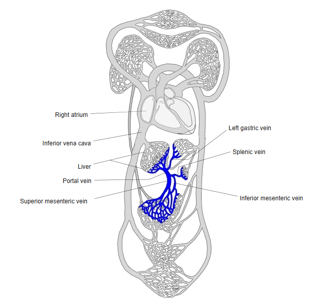
- A short, wide vein that carries blood from the digestive tract, spleen, pancreas and gallbladder to the liver.
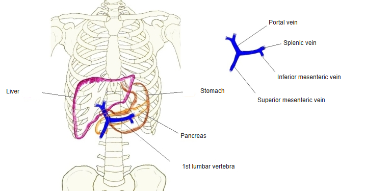
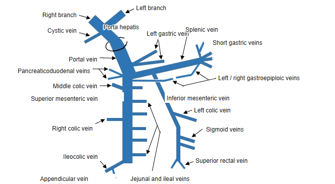
- Formed behind the neck of the pancreas, at about the level of the 1st–2nd lumbar vertebra, by the union of the superior mesenteric vein and the splenic vein.
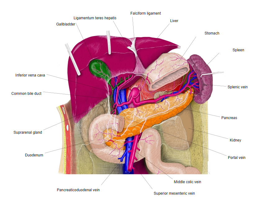
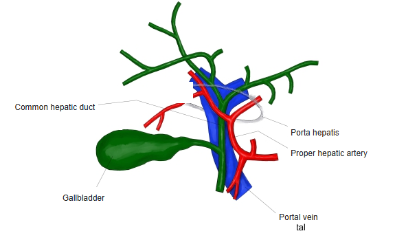
- About 7–8 cm long; runs up behind the 1st part of the duodenum and then in the free edge of the hepatoduodenal ligament to the porta hepatis.
- Blood in it is rich in absorbed nutrients but poor in oxygen; it supplies about 75% of the blood reaching the liver (the rest comes from the hepatic artery).
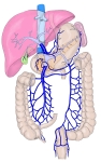
Portal venous system 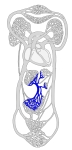
Portal vein and the systemic veins 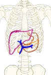
Position of the portal vein
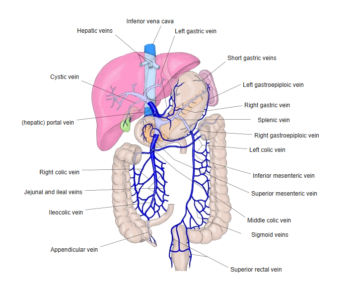
Tributaries
| Vein | Drains |
|---|---|
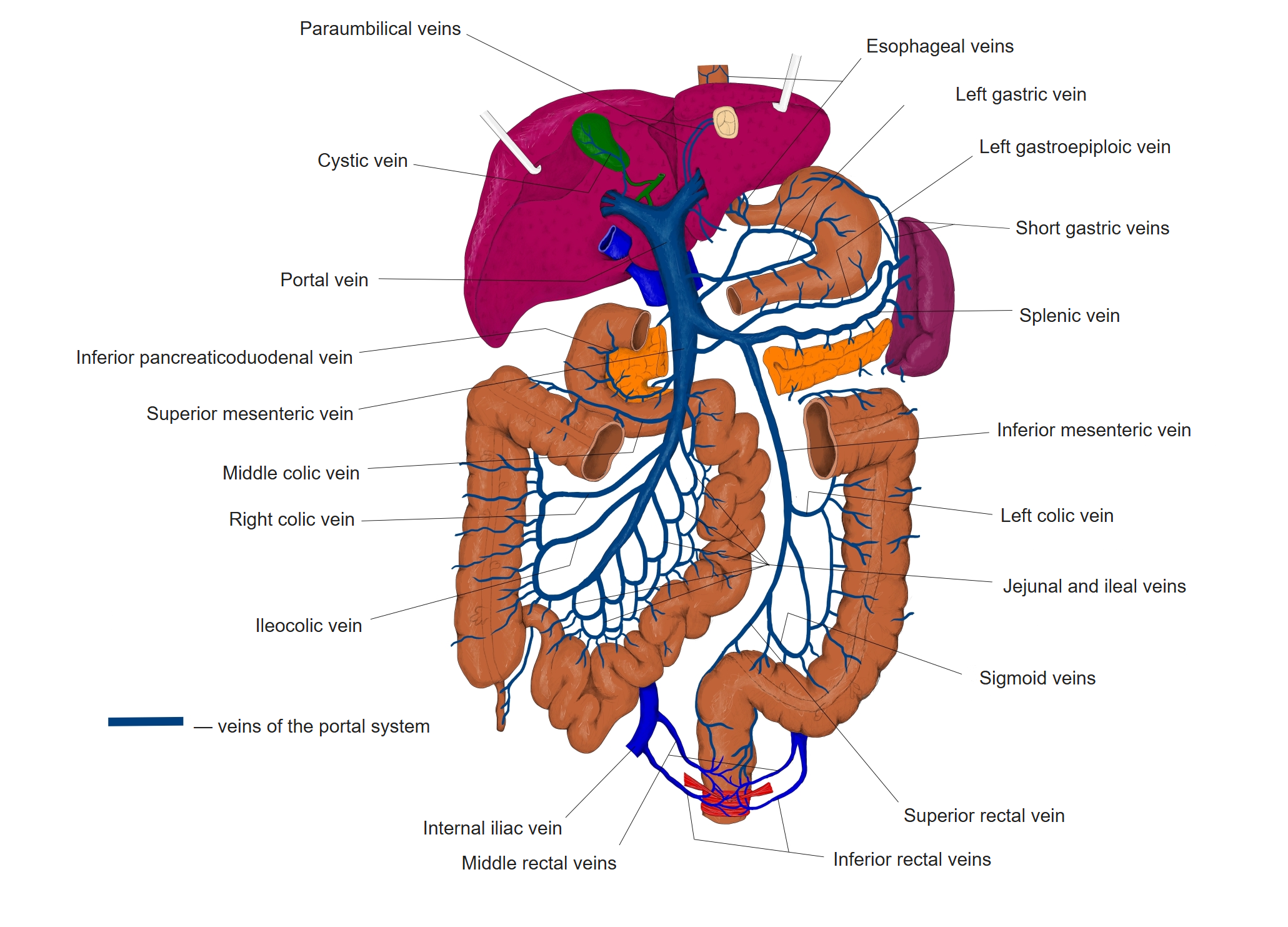
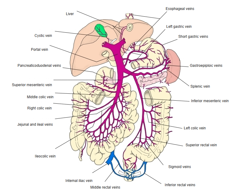
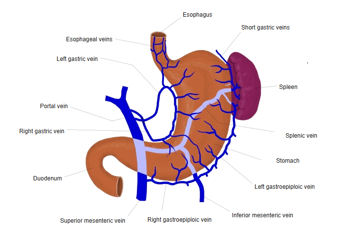
| Splenic vein | Spleen, pancreas, stomach (short gastric and left gastroepiploic veins); usually also the inferior mesenteric vein |
| Superior mesenteric vein | Small intestine, cecum, appendix, ascending and transverse colon, head of pancreas |
| Left gastric vein | Lesser curvature of the stomach and lower esophagus |
| Right gastric vein | Lesser curvature near the pylorus |
| Cystic veins | Gallbladder (may enter the liver directly) |
| Paraumbilical veins | Anterior abdominal wall around the umbilicus (along the round ligament of the liver) |
| Posterior superior pancreaticoduodenal vein | Head of pancreas and duodenum |
Veins of the portal system 
Tributaries of the portal vein (overview) 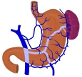
Veins of the stomach and spleen
Course and relations
- Behind it: the inferior vena cava, separated by the omental (epiploic) foramen.
- In the hepatoduodenal ligament it lies behind the proper hepatic artery (left) and the common bile duct (right).
- At the porta hepatis it divides into right and left branches, which follow the segments of the liver and end in the sinusoids through the interlobular veins.
Portal vein and the upper abdominal organs 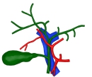
Portal vein at the porta hepatis
Portosystemic anastomoses
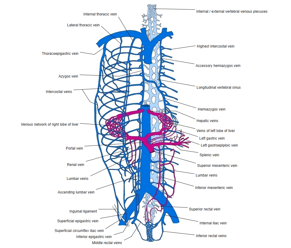
- Where portal and systemic veins meet: lower esophagus (left gastric ↔ esophageal veins of the azygos system), anal canal (superior ↔ middle and inferior rectal veins), umbilicus (paraumbilical ↔ epigastric veins) and the retroperitoneum (colic veins ↔ lumbar and renal veins).
- These channels open up when flow through the liver is blocked.
Veins of the trunk
Clinical notes
- Portal hypertension (most often from cirrhosis) enlarges the anastomoses: esophageal varices (risk of severe bleeding), rectal varices and dilated veins around the umbilicus (caput medusae); it also causes splenomegaly and ascites.
- Portal vein thrombosis can follow cirrhosis, infection or clotting disorders.
References and sources
- Further reading: Portal vein, Wikipedia.