Hepatoduodenal ligament (Latin: ligamentum hepatoduodenale)
- Overview
- Structures passing through it
- Reference websites
- (Wikipedia, websites with illustrations)
- Where it is found
- Related terms
Overview
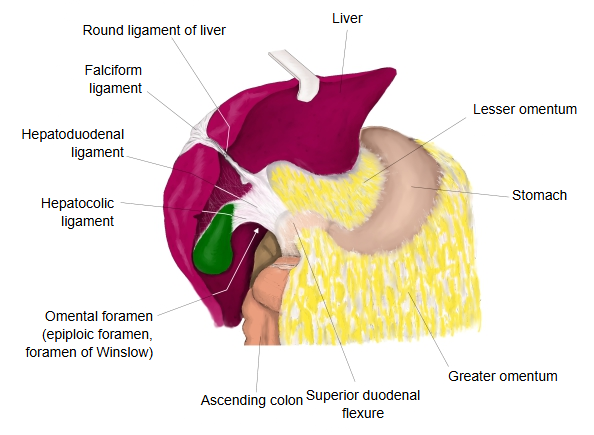
· It is the thickened right free edge of the lesser omentum and carries the portal triad. Its free edge forms the anterior wall of the omental (epiploic) foramen, the opening into the lesser sac. Squeezing the ligament between finger and thumb stops the inflow of blood to the liver during surgery or bleeding (Pringle manoeuvre).
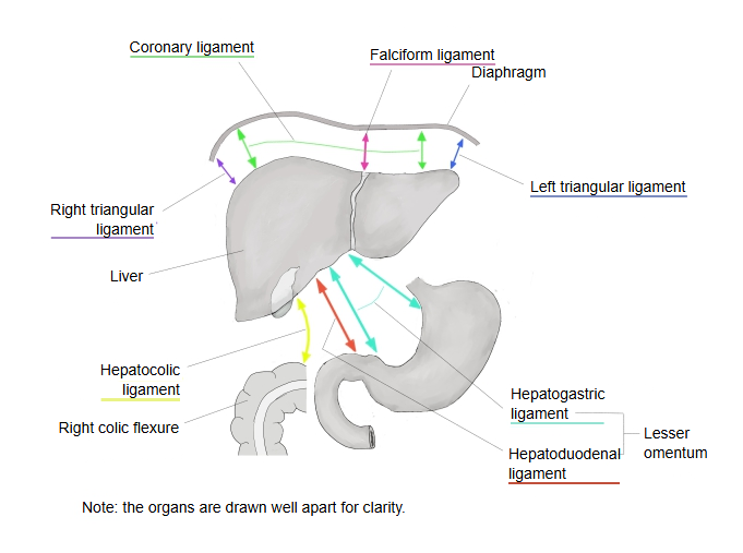
· It develops from the ventral mesogastrium, the mesentery between the stomach and the front of the body wall in the embryo. The part between the liver and the anterior wall becomes the falciform ligament, the reflections onto the diaphragm become the coronary and left and right triangular ligaments, and the part between the liver and the stomach and duodenum becomes the lesser omentum (the hepatogastric and hepatoduodenal ligaments).
- Falciform ligament : from its attachment on the underside of the diaphragm it runs down the front of the liver to the inferior border, and from there to the umbilicus.
- Coronary ligament : two layers of peritoneum, in front and behind, joining the diaphragmatic surface of the liver to the underside of the diaphragm.
- Left and right triangular ligaments : the left and right ends of the coronary ligament.
- Lesser omentum
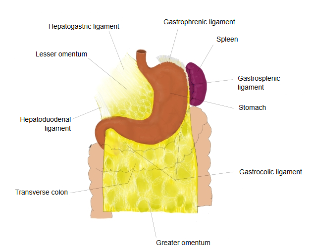
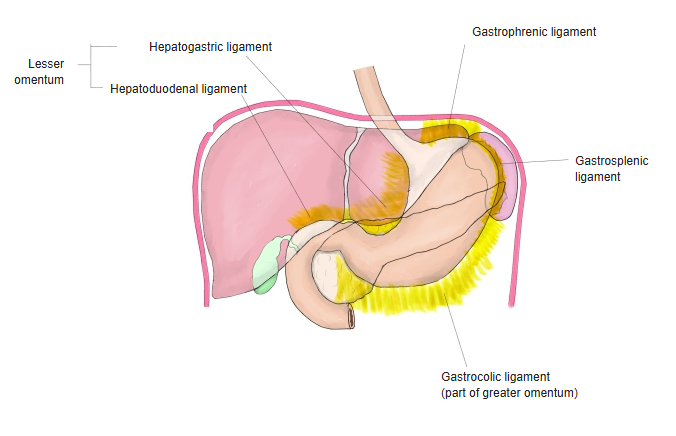
- Hepatogastric ligament : joins the liver (at the porta hepatis) to the lesser curvature of the stomach; forms most of the lesser omentum.
- Hepatoduodenal ligament : joins the liver and the duodenum; the thick right edge of the lesser omentum.
Some textbooks also show the hepatocolic ligament, a variable extension of the lesser omentum to the right colic flexure, as a part of the ventral mesogastrium in their illustrations.
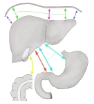 Ligaments of the liver (schematic)
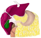 Lesser omentum and omental foramen
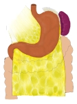 Lesser and greater omenta (anterior)
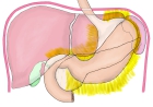 Peritoneal folds around the stomach
Structures passing through it
The following structures run in the hepatoduodenal ligament. The three main ones (the portal triad) lie with the proper hepatic artery in front and to the left, the common bile duct in front and to the right, and the portal vein behind them.
Arteries
1. 1. Proper hepatic artery: the continuation of the common hepatic artery; soon after its origin it runs in the hepatoduodenal ligament. The right gastric artery often arises from it here.
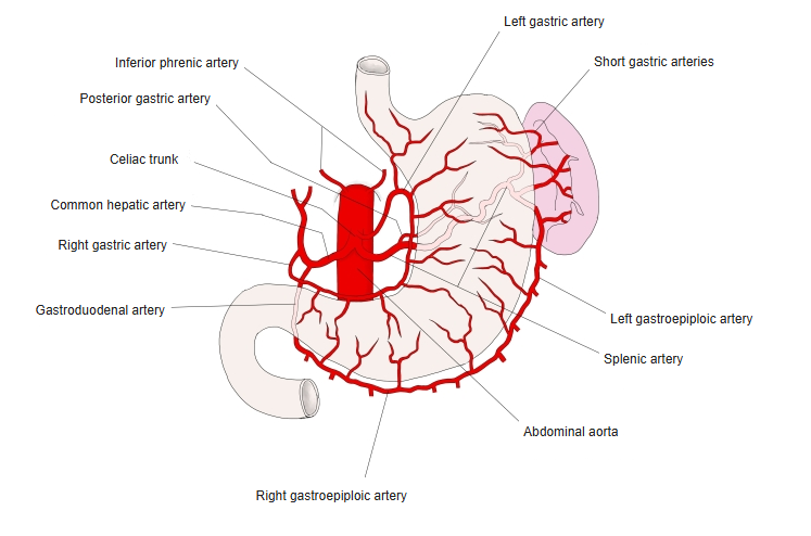
2. Common bile duct (bile duct): runs down in the right free edge of the ligament, in front and to the right of the portal vein.
Veins
1. 1. Portal vein: from where the splenic vein and the superior mesenteric vein join (behind the neck of the pancreas) to where it divides into right and left branches at the porta hepatis.
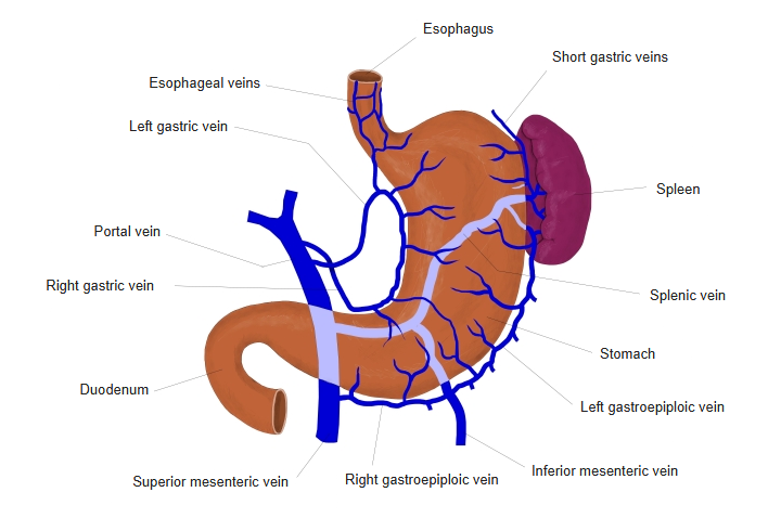
Nerves
1. 1. Hepatic plexus: derived from the celiac plexus and the vagus nerve; it contains sympathetic and parasympathetic fibres.
Lymph vessels / lymph nodes
Lymph vessels from the liver, and hepatic lymph nodes along the hepatic artery, lie in the ligament.
Reference websites
[Wikipedia]
· Hepatoduodenal ligament, Wikipedia.
· Lesser omentum, Wikipedia.
[Websites with illustrations]
· Hepatoduodenal ligament (illustration) (image on cloudfront.net)

· Lesser omentum (illustration) (Pinterest (image))

Where it is found
Related terms
- Lesser omentum
- Hepatogastric ligament
- Portal triad
- Portal vein
- Proper hepatic artery
- Common bile duct
- Porta hepatis
References and sources
- Further reading: Hepatoduodenal ligament, Wikipedia.
- Further reading: Lesser omentum, Wikipedia.
- Federative International Programme for Anatomical Terminology (FIPAT). Terminologia Anatomica, 2nd ed. (TA2), 2019.