Latin: Vena umbilicalis
Overview
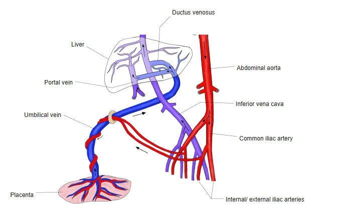
- In the fetus, the vein that carries oxygenated blood from the placenta to the body, through the umbilical cord.
- At first paired; only the left vein persists.
- Enters the abdomen at the umbilicus and runs in the free edge of the falciform ligament to the liver.
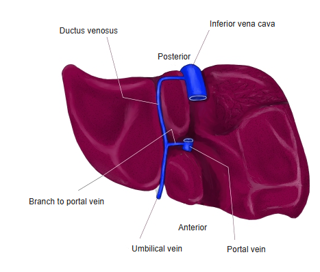
- Most of its blood bypasses the liver through the ductus venosus to the inferior vena cava; the rest goes to the left branch of the portal vein.
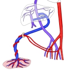
Fetal circulation through the liver
After birth
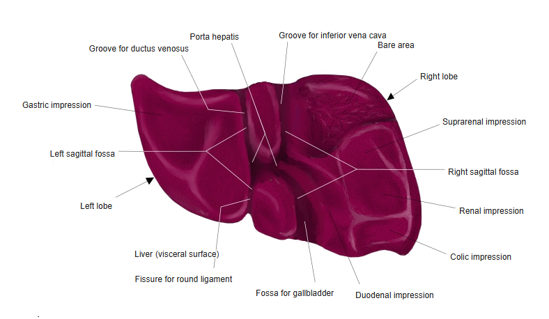
- The umbilical vein closes and becomes the round ligament of the liver (ligamentum teres hepatis).
- The ductus venosus becomes the ligamentum venosum.
Visceral surface of the liver
Clinical notes
- In newborns the umbilical vein can still be catheterized for a few days (umbilical venous catheter).
- It can reopen in portal hypertension (recanalization).
References and sources
- Further reading: Umbilical vein, Wikipedia.