Latin: musculus obliquus inferior bulbi
Overview
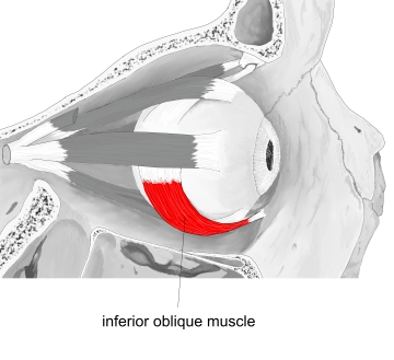
- A thin, narrow extraocular muscle, the shortest of the extraocular muscles (about 35 mm).
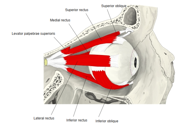
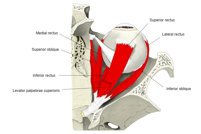
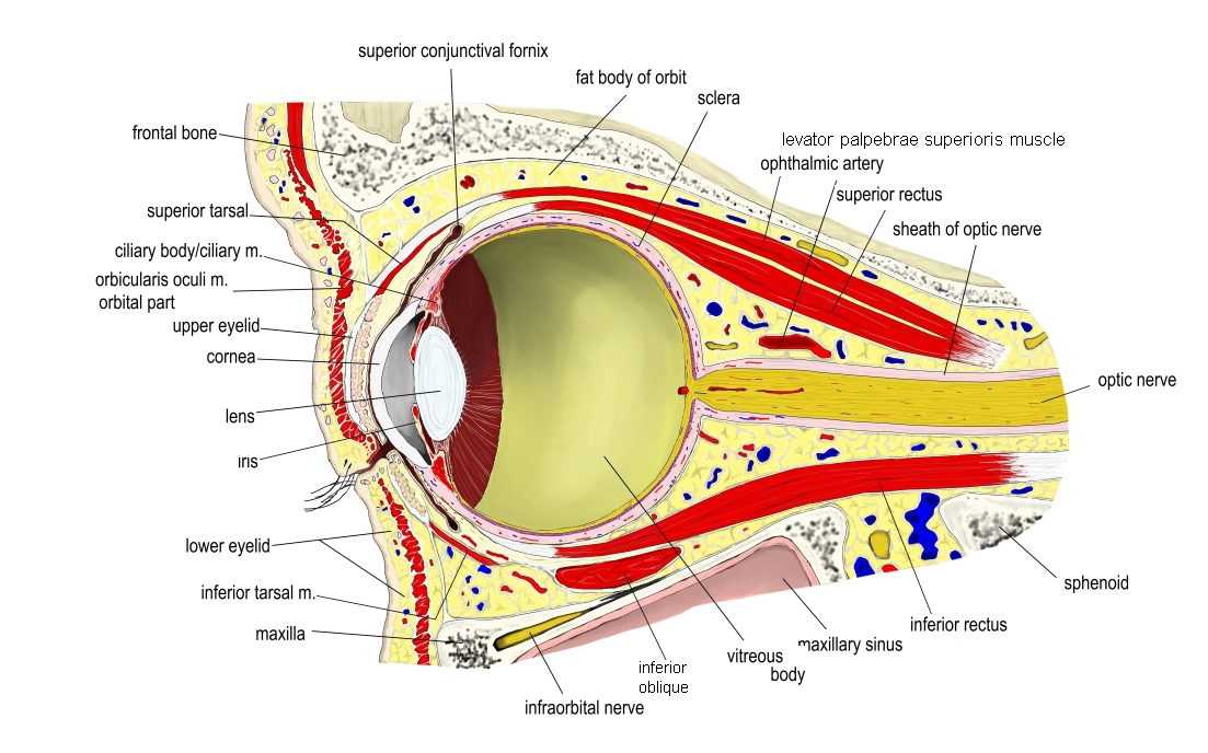
- Unlike the recti, it does not arise from the common tendinous ring but from the front of the orbit.
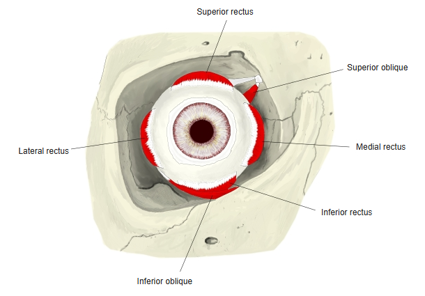
- It passes laterally, backward and upward, first between the inferior rectus and the floor of the orbit, then between the eyeball and the lateral rectus.
Position in the classification
Head › Extraocular muscles
Illustrations
Origin / insertion
- Origin: orbital surface of the maxilla, lateral to the lacrimal groove (nasolacrimal canal), behind the orbital margin
- Insertion: sclera, on the posterolateral part of the eyeball behind the equator, deep to the lateral rectus
Action
- Rotates the eye upward and outward (elevation and abduction)
- Rotates the upper pole of the eye laterally (extorsion) – its primary action
- The only muscle that can elevate the eye when it is fully adducted
Innervation
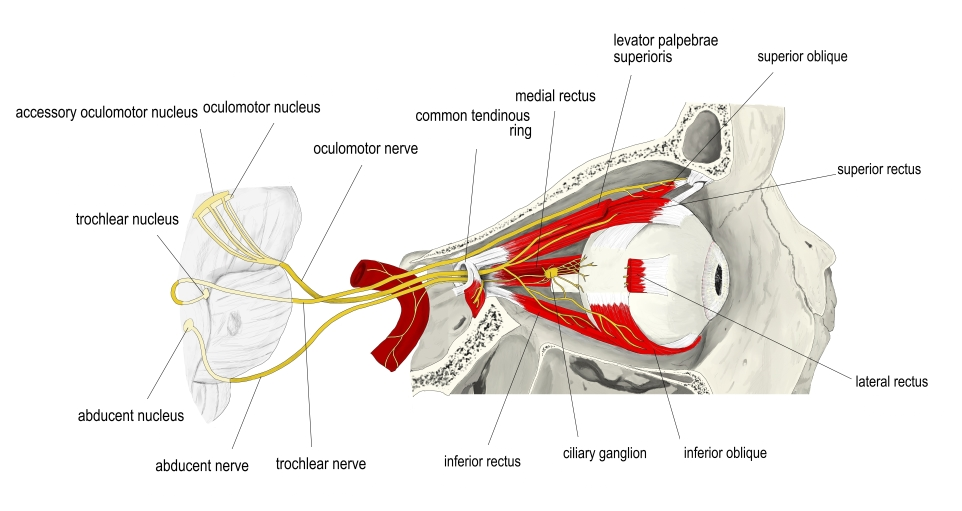
- Oculomotor nerve (cranial nerve III), inferior division
Blood supply
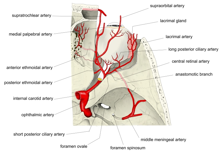
- Ophthalmic artery (muscular branches); also the infraorbital artery
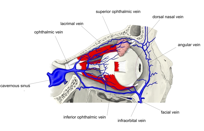
- Venous drainage: ophthalmic veins
Related terms
- Superior oblique
- Inferior rectus
- Lateral rectus
- Oculomotor nerve
- Ophthalmic artery
References and sources
- Further reading: Inferior oblique muscle, Wikipedia.