Latin: musculus extensor digitorum brevis
Overview
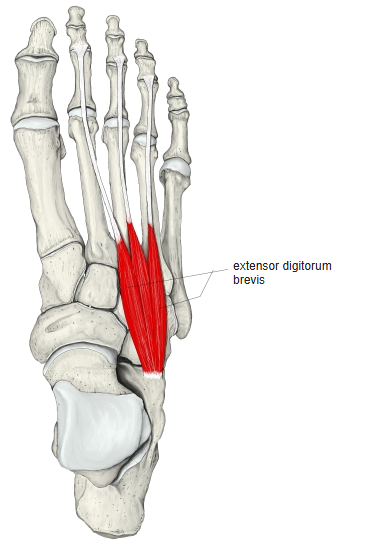
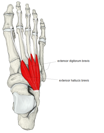
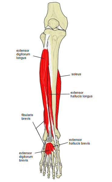
- Arises from the anterior part of the upper and lateral surfaces of the calcaneus and usually divides into two bellies.
- Some anatomists regard it and the neighbouring extensor hallucis brevis as a single muscle.
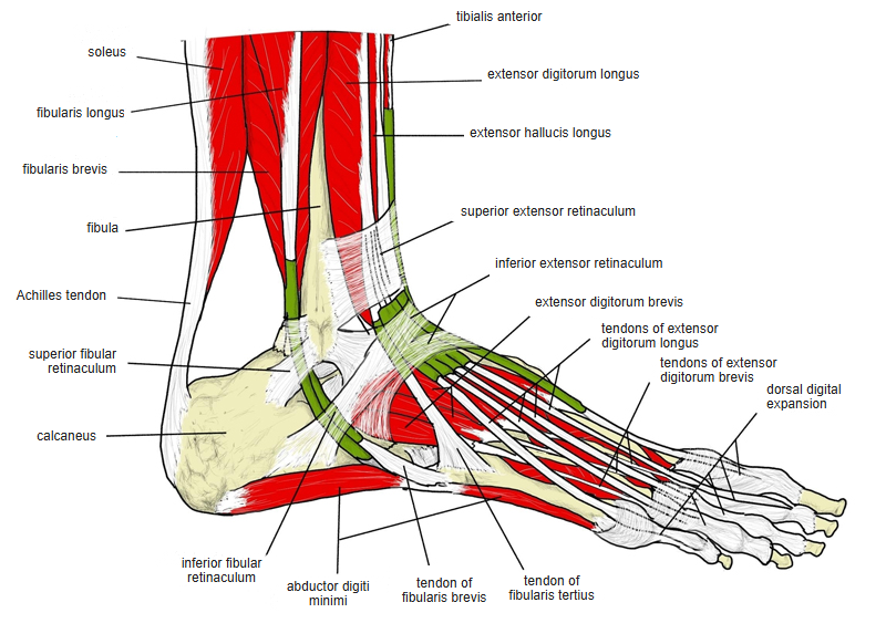
- Its four tendons run forwards and medially: the most medial (the extensor hallucis brevis tendon) inserts into the dorsal surface of the base of the proximal phalanx of the great toe, and the other three join the tendons of the extensor digitorum longus for the 2nd–4th toes.
- In about 8% a tendon also goes to the 5th toe.
- Its action is most noticeable when the foot is dorsiflexed and the long extensor tendons are relaxed.
Position in the classification
Lower Limb › Foot › Dorsum of foot
- Extensor digitorum brevis
- Extensor hallucis brevis
Illustrations
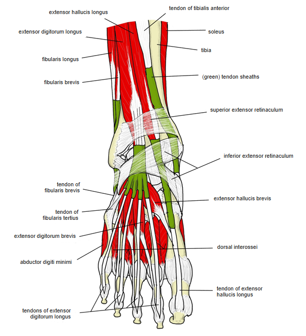
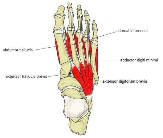
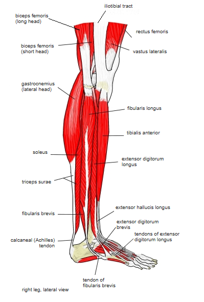
Origin / insertion
- Origin: calcaneus (the anterior part of its upper and lateral surfaces); also the lateral talocalcaneal ligament and the stem of the inferior extensor retinaculum
- Insertion: dorsal digital expansions of the 2nd–4th toes; each tendon joins the lateral side of the corresponding tendon of the extensor digitorum longus
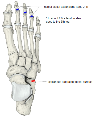
Action
- Extends the 2nd–4th toes, drawing them upwards and laterally
Innervation
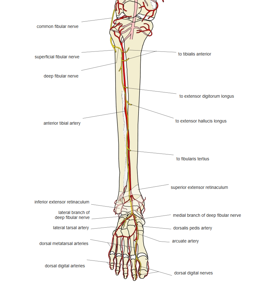
- Deep fibular nerve (L5, S1; some sources L4–S1)
Blood supply
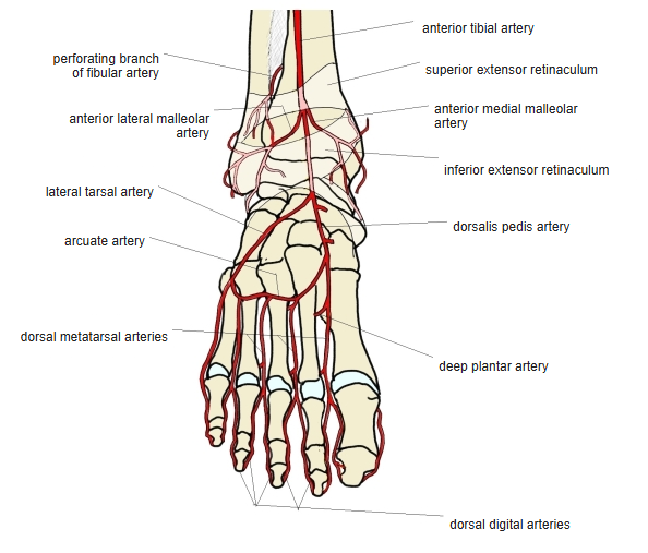
- Lateral tarsal artery (from the dorsalis pedis artery), perforating branch of the fibular artery
Related terms
- Extensor hallucis brevis
- Extensor digitorum longus
- Calcaneus
- Inferior extensor retinaculum
- Deep fibular nerve
- Dorsalis pedis artery
- Lateral tarsal artery
References and sources
- Further reading: Extensor digitorum brevis muscle, Wikipedia.