Intercellular bridge
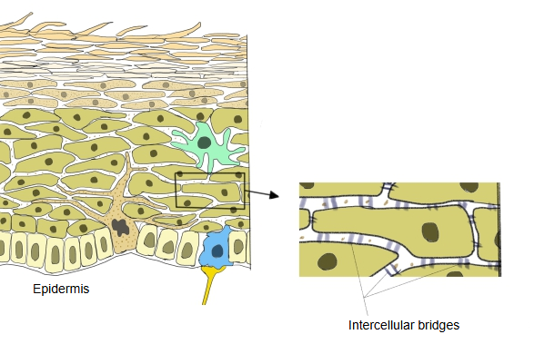
Intercellular bridges are very fine connections between neighbouring flattened (squamous) epithelial cells, seen with the light microscope.
Cells of the stratified squamous epithelium of the epidermis and of the mucous membranes of the oral cavity, oesophagus and other parts
They are most obvious in the stratum spinosum of the epidermis. With the electron microscope each bridge turns out to be a pair of desmosomes: the two cell membranes stay separate, held together by attachment plaques into which keratin filaments are anchored. In ordinary sections the cells shrink apart and remain attached only at these points, which makes the keratinocytes look covered in spines (prickle cells). The bridges hold the cells firmly together, so that the epithelium resists stretching and rubbing. Sources differ: the same term is also used for true cytoplasmic bridges, for example those that link developing sperm cells.
Reference websites
[Wikipedia]
· Desmosome, Wikipedia.
· Stratum spinosum, Wikipedia.
Related terms
References and sources
- Further reading: Desmosome, Wikipedia.
- Further reading: Stratum spinosum, Wikipedia.
- Federative International Programme for Anatomical Terminology (FIPAT). Terminologia Anatomica, 2nd ed. (TA2), 2019.