Latin: musculus masseter
Overview
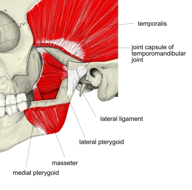
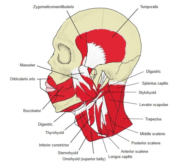
- One of the four muscles of mastication and the most superficial of them; a thick, somewhat quadrilateral muscle.
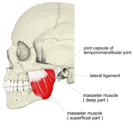
- It has two layers, a superficial part and a deep part, whose fibres run in different directions; in front, the deep part is partly covered by the superficial part.
- Superficial part: wider, with a thick belly and a strong tendon.
- Deep part: smaller, but with a higher proportion of muscle fibres.
- It can be seen and felt under the skin when the teeth are clenched.
- Its posterior part is covered by the parotid gland; the zygomaticus major and minor sometimes cross near the origin of the superficial part.
Position in the classification
Head › Muscles of mastication
Illustrations
Origin / insertion
- Origin:
- Superficial part: zygomatic process of the maxilla and the anterior two-thirds of the lower border of the zygomatic arch (by a thick tendinous aponeurosis); runs obliquely downward and backward
- Deep part: middle and posterior parts of the zygomatic arch and its medial surface; runs almost vertically downward
- Insertion: the masseteric tuberosity on the outer surface of the ramus and angle of the mandible
- Superficial part: angle and lower half of the lateral surface of the ramus
- Deep part: upper half of the ramus and the lateral surface of the coronoid process
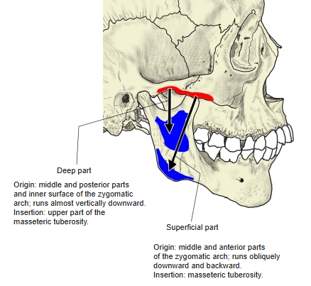
Action
- Raises the mandible and brings the teeth together (closes the jaw)
- The superficial fibres can also help to protrude the jaw.
Innervation
- Masseteric nerve, a branch of the mandibular nerve (V3)
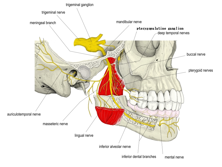
Blood supply
- Masseteric artery, a branch of the maxillary artery
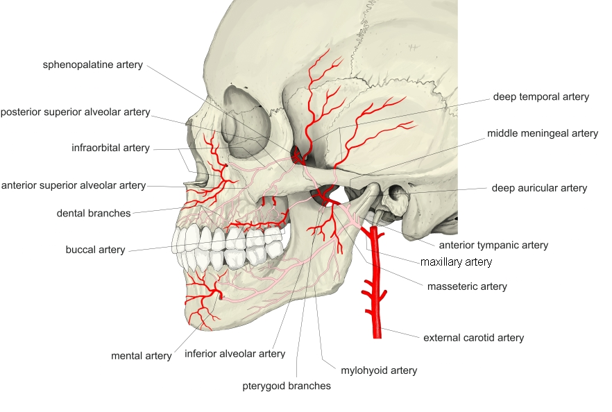
Related terms
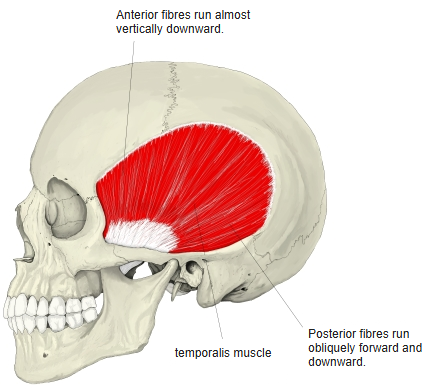
- Muscles of mastication
- Temporalis
- Medial pterygoid
- Lateral pterygoid
- Zygomatic arch
- Parotid gland
- Mandibular nerve
References and sources
- Further reading: Masseter muscle, Wikipedia.
- Gray H. Anatomy of the Human Body, 20th ed. (1918), “4e. The Muscles of Mastication”, Bartleby.com.