Medial pterygoid (Latin: musculus pterygoideus medialis)
- Overview
- Illustrations
- Origin / insertion
- Action
- Nerve / blood supply
- Reference websites
- (Wikipedia, websites with illustrations)
- Related terms
Overview
- Facial muscles (24)
- Muscles of mastication (4) : masseter, temporalis, lateral pterygoid, medial pterygoid
- Extra-ocular muscles (7)
- Muscles of the auditory ossicles (2)
- Muscles of the tongue (7)
- Muscles of the soft palate and fauces (5)
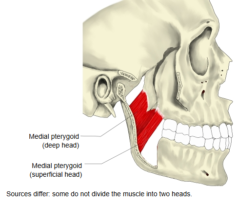
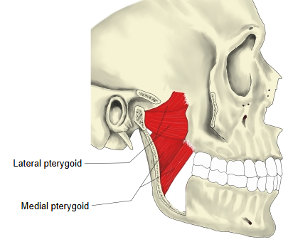
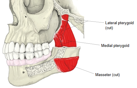
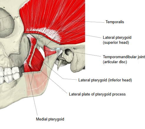
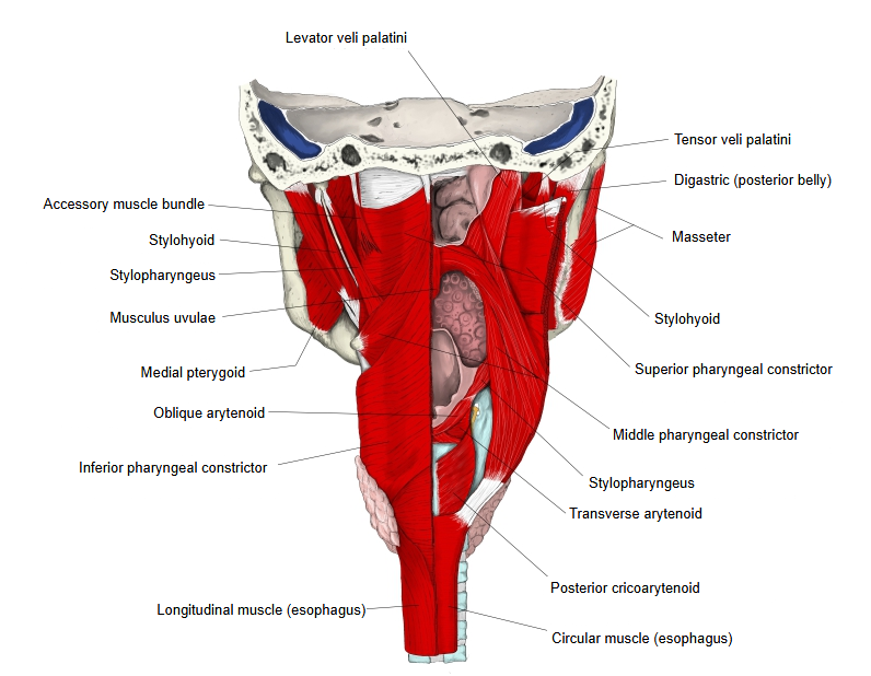
· A thick, four-sided muscle on the inner side of the ramus of the mandible. It runs obliquely from front and above to back and below.
· Most texts describe two heads: a large deep head and a small superficial head. Some texts do not divide it.
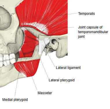
· On the outer side of the ramus lies the masseter. The two muscles pull in the same direction (they are synergists) and form a sling around the angle of the mandible.
Illustrations
More figures: Multi-muscle illustrations: Head · Muscles of the head (list)
Origin / insertion
[Origin] : two heads.
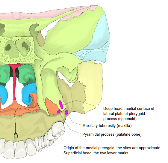
Deep head (the larger part): the medial surface of the lateral plate of the pterygoid process of the sphenoid bone, in the pterygoid fossa, and the pyramidal process of the palatine bone.
Superficial head (small): the maxillary tuberosity of the maxilla and the pyramidal process of the palatine bone. It lies on the outer side of the lower head of the lateral pterygoid.
The fibres run downward, backward and laterally.
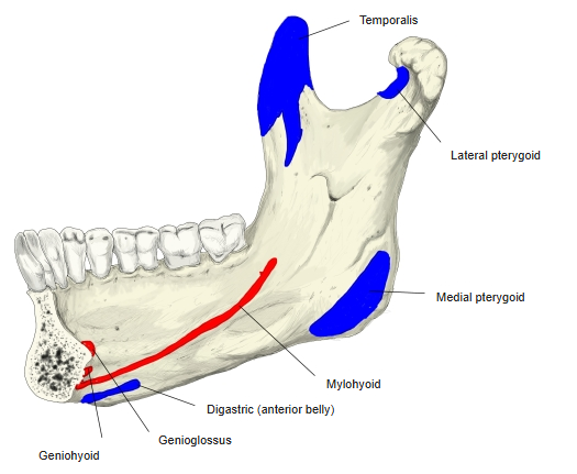
[Insertion] : the pterygoid tuberosity: the rough lower and back part of the medial surface of the ramus and the angle of the mandible, up to the level of the mandibular foramen.
 Origin
 Insertion
Action
Elevates the mandible (closes the mouth), with the masseter and temporalis. With the lateral pterygoid it protrudes the mandible. Acting on one side, it pulls the mandible toward the opposite side; the right and left muscles acting in turn produce the grinding movements of chewing.
Nerve / blood supply
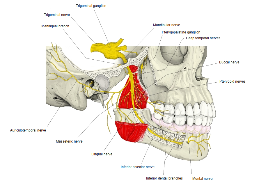
· Nerve : nerve to medial pterygoid, a branch of the main trunk of the mandibular nerve (third division of the trigeminal nerve, cranial nerve V), given off before the nerve divides. The other muscles of mastication are supplied from the anterior division.
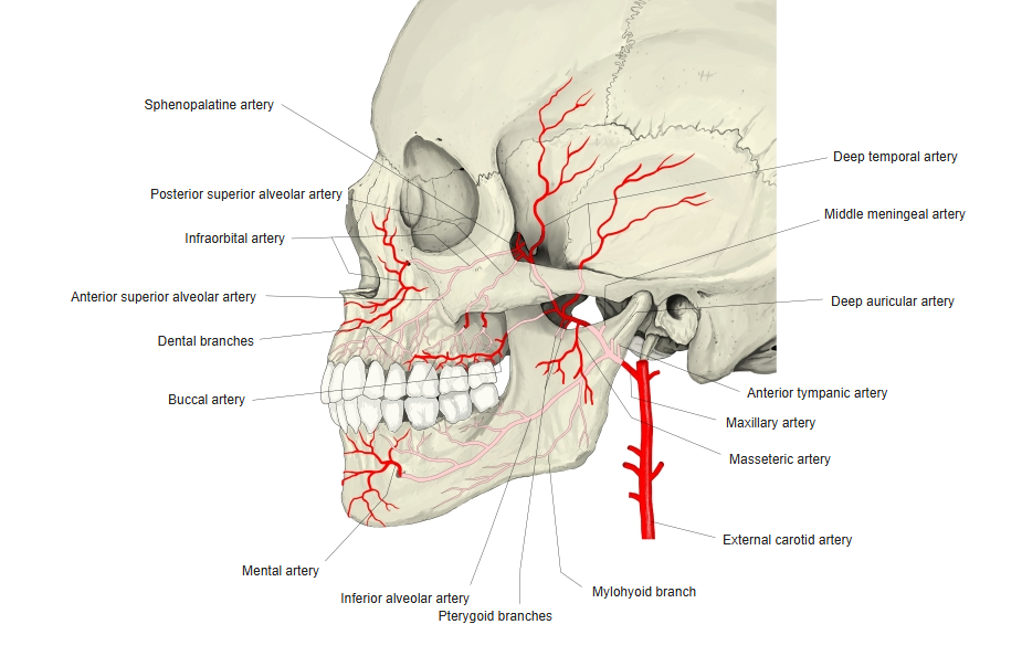
· Arteries : pterygoid branches of the maxillary artery (one of the two end branches of the external carotid artery); small branches of the facial artery also reach it.
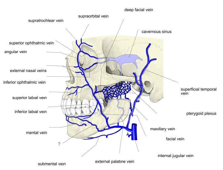
· Veins : to the pterygoid plexus, which drains through the maxillary veins.
 Mandibular nerve
 Maxillary artery
 Veins of the face
Reference websites
[Wikipedia]
· Medial pterygoid muscle, Wikipedia.
[Websites with illustrations]
· Medial pterygoid (Kenhub)
Related terms
- Pterygoid fossa
- Pterygoid process
- Maxillary tuberosity
- Pterygoid tuberosity
- Temporomandibular joint
- Lateral pterygoid
- Masseter
- Temporalis
- Mandibular nerve
References and sources
- FIPAT. Terminologia Anatomica, 2nd ed. (TA2), 2019: musculus pterygoideus medialis.
- Medial pterygoid muscle, Wikipedia.