← Bone illustrations: Skull — Parts of the skull
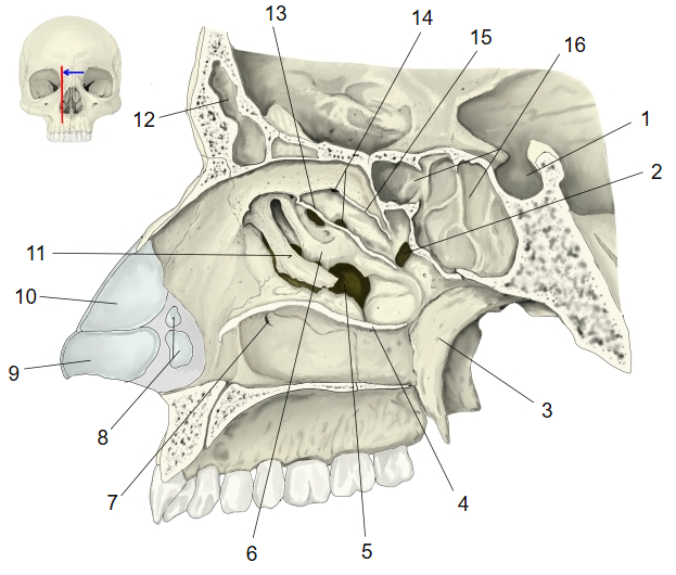
| No. | Name | Page |
|---|---|---|
| 1 | Hypophysial fossa | — |
| 2 | Sphenopalatine foramen | — |
| 3 | Sphenoid bone, pterygoid process, medial plate | Detail |
| 4 | Inferior nasal concha (cut surface) | Detail |
| 5 | Maxillary hiatus | — |
| 6 | Ethmoid bone, ethmoidal bulla | Detail |
| No. | Name | Page |
|---|---|---|
| 7 | Opening of the nasolacrimal duct | — |
| 8 | Minor alar cartilages | — |
| 9 | Major alar cartilage | — |
| 10 | Lateral nasal cartilage | — |
| 11 | Ethmoid bone, uncinate process | Detail |
| 12 | Frontal sinus | — |
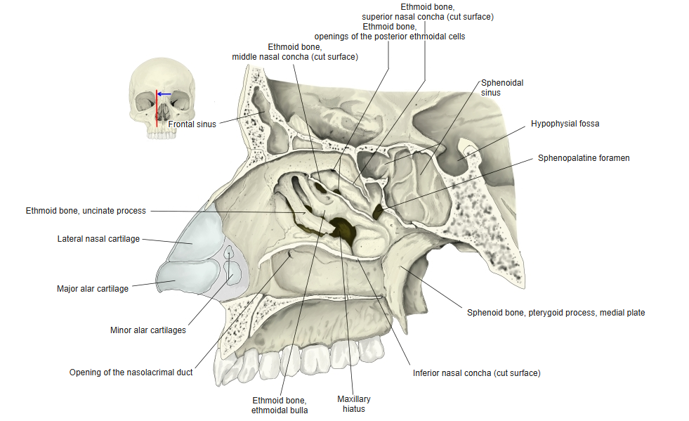
More illustrations: Skull — Parts of the skull
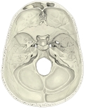 Cranial base (internal surface)
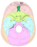 Cranial base (internal surface, bones colour-coded)
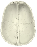 Calvaria (internal surface)
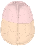 Calvaria (internal surface, bones colour-coded)
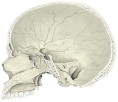 Skull (sagittal section)
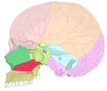 Skull (sagittal section, bones colour-coded)
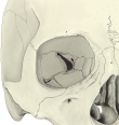 Right orbit
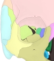 Right orbit (bones colour-coded)
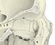 Right orbital opening, section (lateral view)
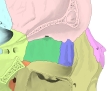 Right orbital opening, section (lateral view, bones colour-coded)
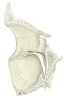 Right orbital opening, section (medial view)
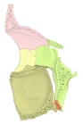 Right orbital opening, section (medial view, bones colour-coded)
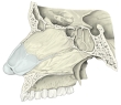 Nasal septum, sagittal section (from the left)
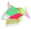 Nasal septum, sagittal section (from the left, bones colour-coded)
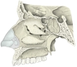 Lateral wall of the nasal cavity (from the left)
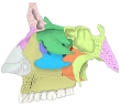 Lateral wall of the nasal cavity (from the left, bones colour-coded)
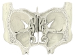 Orbits and nasal cavity (coronal section)
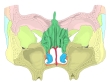 Orbits and nasal cavity (coronal section, bones colour-coded)
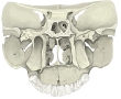 Orbit to palate (coronal section)
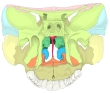 Orbit to palate (coronal section, bones colour-coded)
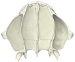 Skull, transverse section (lower nasal cavity)
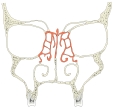 Nasal cavity and paranasal sinuses (coronal section)