Latin: os sphenoidale
Overview
- An unpaired bone in the middle of the cranial base, shaped like a butterfly or a bat with spread wings.
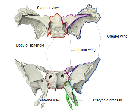
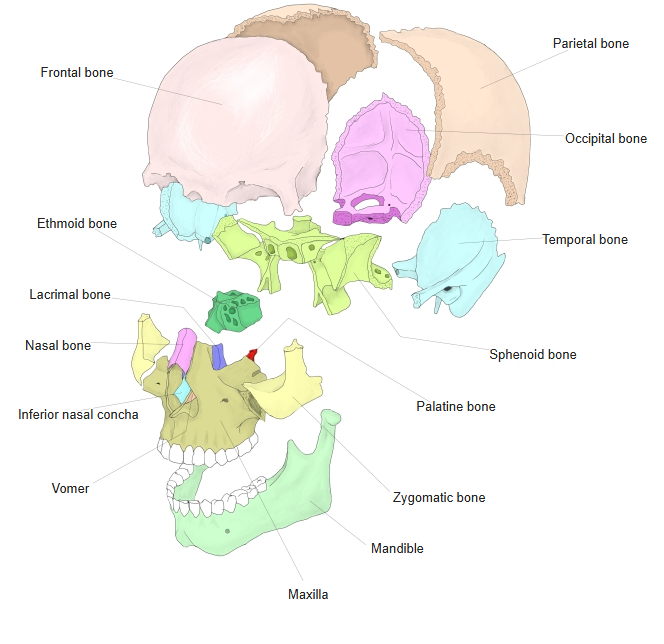
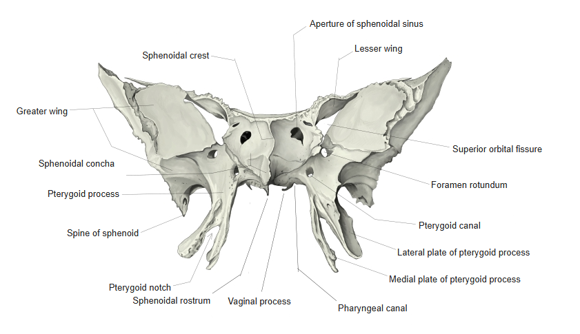
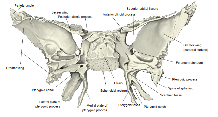
- Its parts are the body (containing the sphenoidal sinus, with the sella turcica above), the paired greater wings and lesser wings, and the paired pterygoid processes (each with a medial and lateral plate) that project downwards.
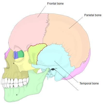
- It meets almost all the other bones of the cranium: the frontal, parietal, temporal, occipital and ethmoid bones, and the vomer, zygomatic and palatine bones.
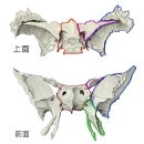
Parts of the sphenoid
- Bones of the skull
- Neurocranium (cranial bones)
- Frontal bone
- Parietal bone (2)
- Occipital bone
- Temporal bone (2)
- Sphenoid bone
- Ethmoid bone
- Viscerocranium (facial bones)
- Nasal bone (2)
- Vomer
- Lacrimal bone (2)
- Inferior nasal concha (2)
- Maxilla (2)
- Zygomatic bone (2)
- Palatine bone (2)
- Mandible
- Hyoid bone
- Neurocranium (cranial bones)
Illustrations
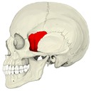
Skull (left lateral) 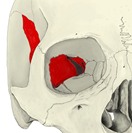
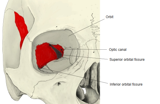
Left orbit 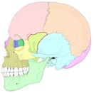
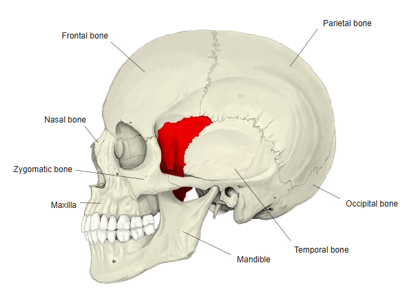
Skull (left lateral, coloured) 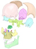
Bones of the skull (separated) 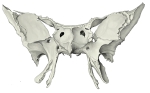
Sphenoid (anterior) 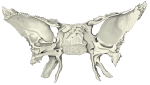
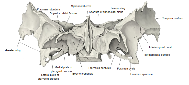
Sphenoid (posterior) 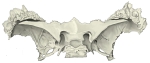
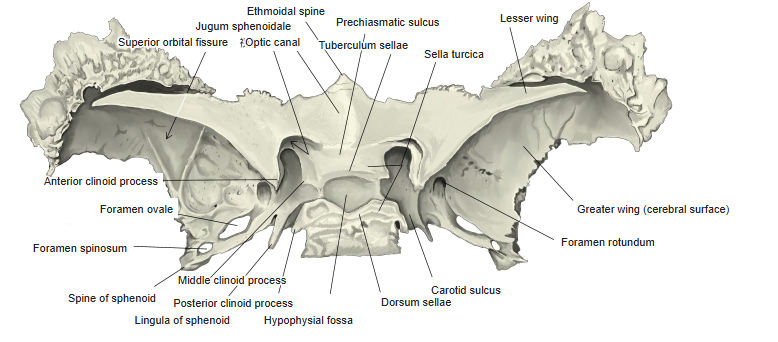
Sphenoid (from above) 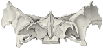
Sphenoid (from below)
Articulations
| # | Bone | Joint |
|---|---|---|
| 1 | Frontal bone | Sphenofrontal suture |
| 2 | Parietal bones | Sphenoparietal suture |
| 3 | Temporal bones | Sphenosquamous suture, sphenopetrosal fissure |
| 4 | Occipital bone | Spheno-occipital synchondrosis (fuses in the adult) |
| 5 | Ethmoid bone | Sphenoethmoidal suture |
| 6 | Vomer | Sphenovomerine suture |
| 7 | Zygomatic bones | Sphenozygomatic suture |
| 8 | Palatine bones | Pterygoid processes and body |
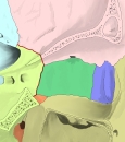
Medial wall of the right orbit 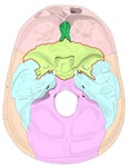
Internal cranial base 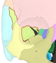
Coronal section of the skull 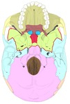
External cranial base 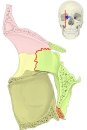
Sagittal section through the orbit
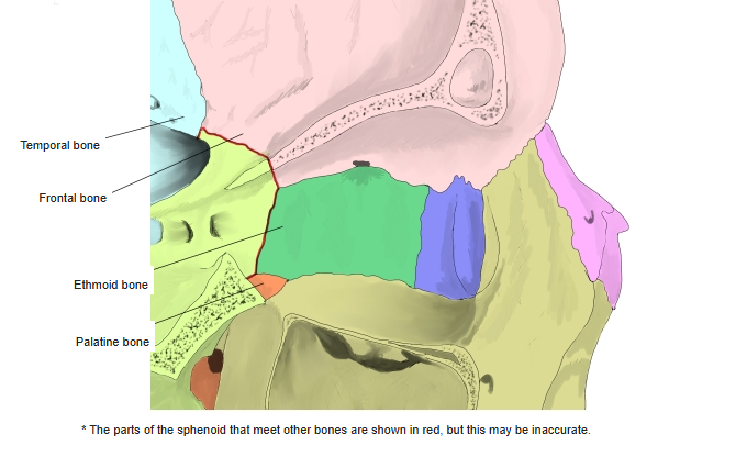
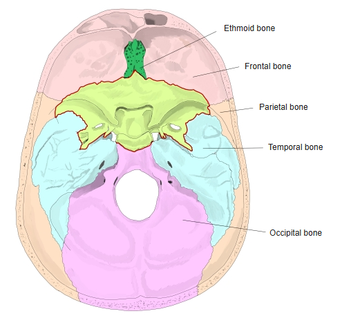
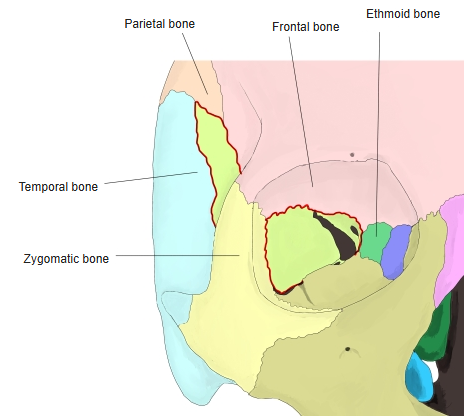
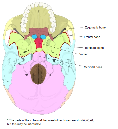
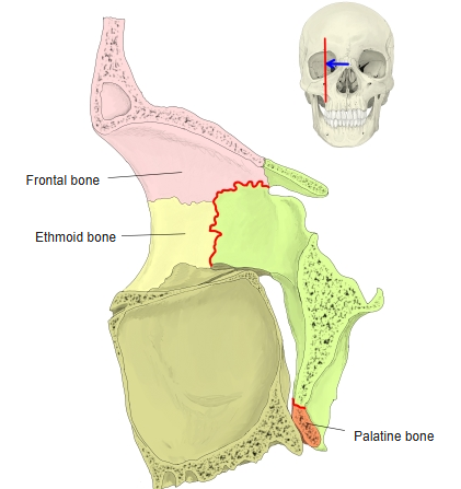
In these figures the parts of the sphenoid that meet other bones are shown in red; the extent shown may be slightly inaccurate.
Ligaments
The following ligaments attach to the sphenoid bone. The list is intended to be comprehensive but may omit minor ligaments.
| # | Ligament | Connects |
|---|---|---|
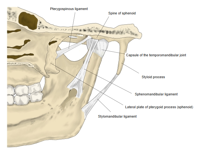
| 1 | Pterygospinous ligament | Spine of sphenoid – lateral plate of pterygoid process |
| 2 | Sphenomandibular ligament | Spine of sphenoid – lingula of mandible |
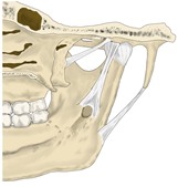
Ligaments of the temporomandibular joint (medial)
Muscle attachments
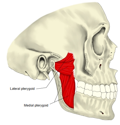
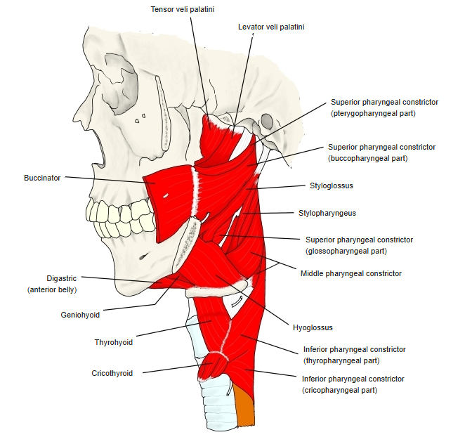
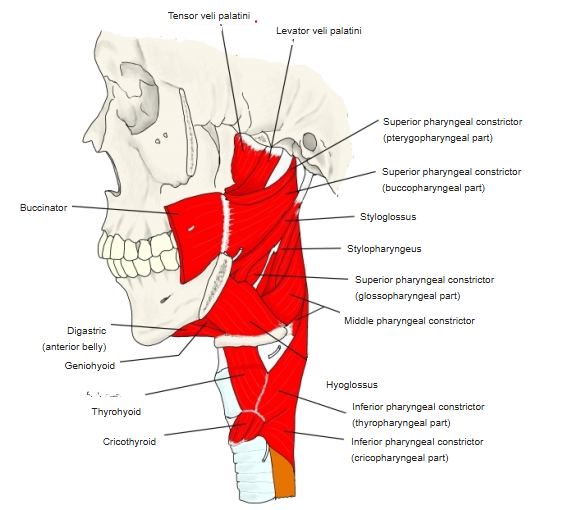
- Origin of: Temporalis, Lateral pterygoid, Medial pterygoid, Tensor veli palatini, Superior pharyngeal constrictor, Levator veli palatini
- Insertion of: none
Temporalis arises partly from the temporal surface of the greater wing; lateral pterygoid from the infratemporal surface and the lateral plate; medial pterygoid from the medial surface of the lateral plate; the superior pharyngeal constrictor from the pterygoid hamulus.
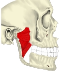
Pterygoid muscles 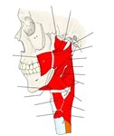
Muscles of the pharynx and soft palate (lateral) 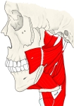
Muscles of the pharynx (lateral)
Main features
- Body – contains the sphenoidal sinus, opening in front through the apertures of the sphenoidal sinus beside the sphenoidal crest. On top lies the sella turcica: the tuberculum sellae in front, the hypophysial fossa (for the pituitary gland) in the middle and the dorsum sellae behind, with the anterior, middle and posterior clinoid processes.
- Lesser wings – triangular plates at the front of the body; each contains the optic canal at its root and ends in the anterior clinoid process.
- Greater wings – with cerebral, temporal, infratemporal and orbital surfaces; they carry the foramen rotundum, foramen ovale and foramen spinosum, and end behind in the spine of the sphenoid.
- Superior orbital fissure – the gap between the lesser and greater wings.
- Pterygoid processes – each with a medial plate (ending in the pterygoid hamulus) and a lateral plate, enclosing the pterygoid fossa; the pterygoid canal runs through their root.
References and sources
- Further reading: Sphenoid bone, Wikipedia.