Latin: os nasale
Overview
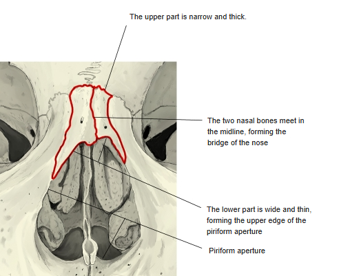
- A small paired bone of the facial skeleton that forms the root and bridge of the nose.
- Roughly trapezoid in shape: the upper part is narrow and thick, the lower part wide and thin. The left and right bones meet in the midline; they are often unequal in size.
- Above, it meets the frontal bone; below, its free edge forms the upper margin of the piriform aperture.
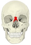
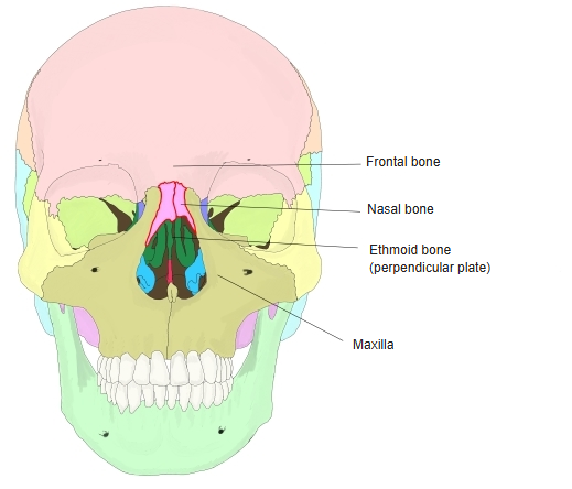
Skull (anterior) 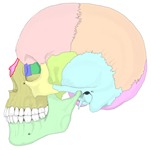
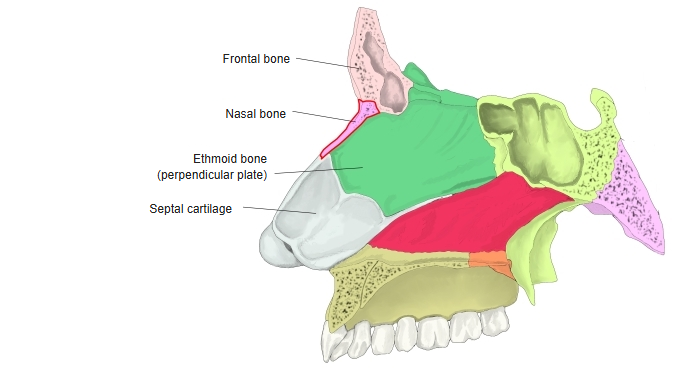
Skull (left lateral) 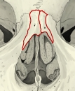
Shape of the nasal bones
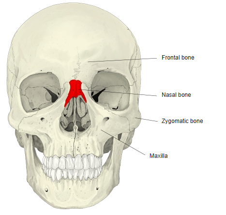
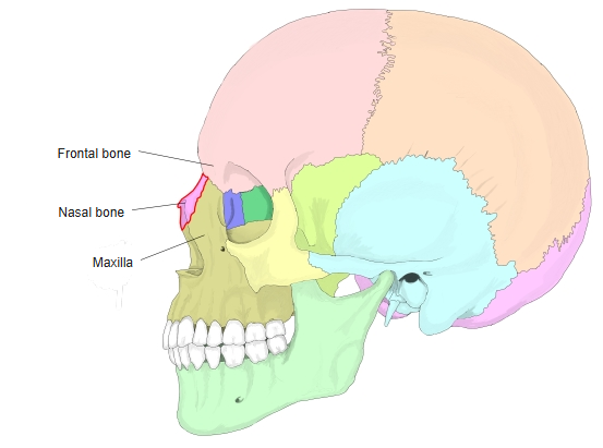
- Bones of the skull
- Neurocranium (cranial bones)
- Frontal bone
- Parietal bone (2)
- Occipital bone
- Temporal bone (2)
- Sphenoid bone
- Ethmoid bone
- Viscerocranium (facial bones)
- Nasal bone (2)
- Vomer
- Lacrimal bone (2)
- Inferior nasal concha (2)
- Maxilla (2)
- Zygomatic bone (2)
- Palatine bone (2)
- Mandible
- Hyoid bone
- Neurocranium (cranial bones)
Illustrations
Articulations
| # | Bone | Joint |
|---|---|---|
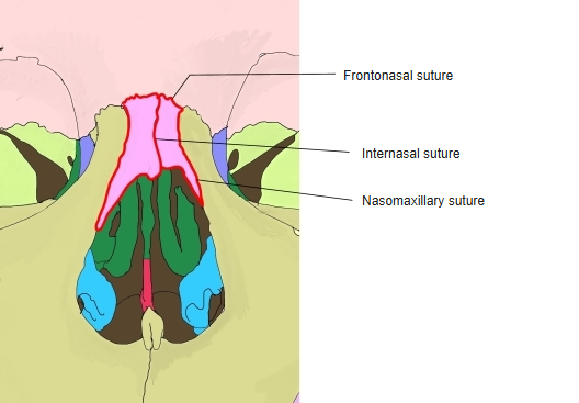
| 1 | Frontal bone | Frontonasal suture |
| 2 | Opposite nasal bone | Internasal suture |
| 3 | Maxilla (frontal process) | Nasomaxillary suture |
| 4 | Ethmoid bone (perpendicular plate) | Behind the internasal suture |
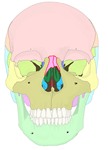
Neighbouring bones (anterior) 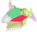
Neighbouring bones (sagittal section) 
Sutures of the nasal bones
Muscle attachments
- Origin of: Procerus
- Insertion of: none
Procerus arises from the fascia over the lower part of the nasal bone and the upper part of the lateral nasal cartilage.
Borders
- Superior border – thick and serrated; joins the nasal part of the frontal bone (frontonasal suture).
- Inferior border – thin and free; forms the upper edge of the piriform aperture and usually has a notch.
- Medial border – joins the opposite nasal bone (internasal suture); behind, it rests on the nasal spine of the frontal bone and the perpendicular plate of the ethmoid.
- Lateral border – joins the frontal process of the maxilla (nasomaxillary suture).
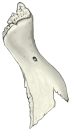
Borders of the right nasal bone
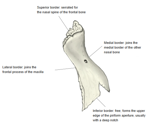
Surfaces
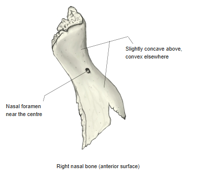
- Anterior (outer) surface – slightly concave above and convex below; a small nasal foramen for a vein usually lies near its centre.
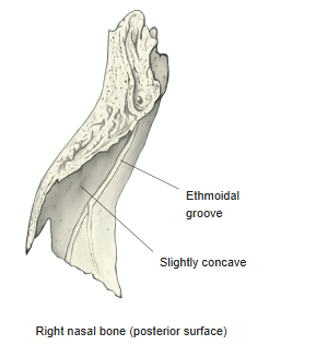
- Posterior (inner) surface – faces the nasal cavity and is slightly concave; it carries the ethmoidal groove for the external nasal branch of the anterior ethmoidal nerve.
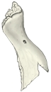
Right nasal bone (anterior surface) 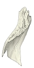
Right nasal bone (posterior surface)
References and sources
- Further reading: Nasal bone, Wikipedia.