Latin: os frontale
Overview
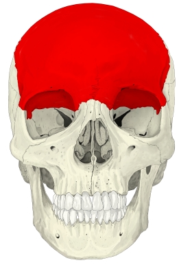
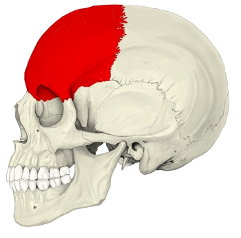
- One of the bones of the neurocranium; it forms the forehead, the front of the vault and most of the roof of the orbits.
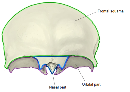
- It has three parts: the frontal squama, the paired orbital parts and the nasal part.
- It meets 12 bones of 7 kinds: the parietal bones (2), sphenoid, ethmoid, nasal bones (2), maxillae (2), lacrimal bones (2) and zygomatic bones (2).
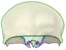
Parts of the frontal bone
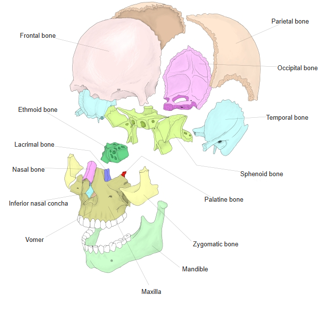
- Bones of the skull
- Neurocranium (cranial bones)
- Frontal bone
- Parietal bone (2)
- Occipital bone
- Temporal bone (2)
- Sphenoid bone
- Ethmoid bone
- Viscerocranium (facial bones)
- Nasal bone (2)
- Vomer
- Lacrimal bone (2)
- Inferior nasal concha (2)
- Maxilla (2)
- Zygomatic bone (2)
- Palatine bone (2)
- Mandible
- Hyoid bone
- Neurocranium (cranial bones)
Illustrations
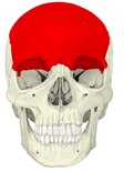
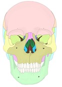
Skull (anterior) 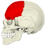
Skull (left lateral) 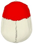
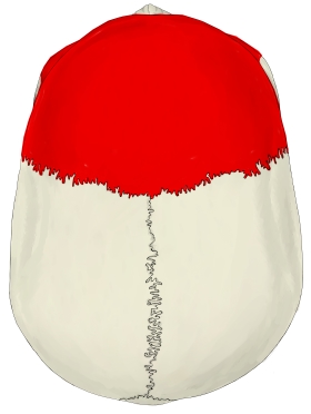
Skull (from above) 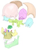
Bones of the skull (separated)
Articulations
| # | Bone | Joint |
|---|---|---|
| 1 | Parietal bones (2) | Coronal suture |
| 2 | Sphenoid | Sphenofrontal suture |
| 3 | Ethmoid | Frontoethmoidal suture |
| 4 | Nasal bones (2) | Frontonasal suture |
| 5 | Maxillae (2) | Frontomaxillary suture |
| 6 | Lacrimal bones (2) | Frontolacrimal suture |
| 7 | Zygomatic bones (2) | Frontozygomatic suture |
Neighbouring bones (anterior) 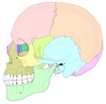
Neighbouring bones (left lateral) 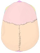
Neighbouring bones (from above) 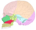
Neighbouring bones (median section) 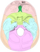
Neighbouring bones (internal cranial base) 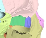
Neighbouring bones (medial wall of the right orbit)
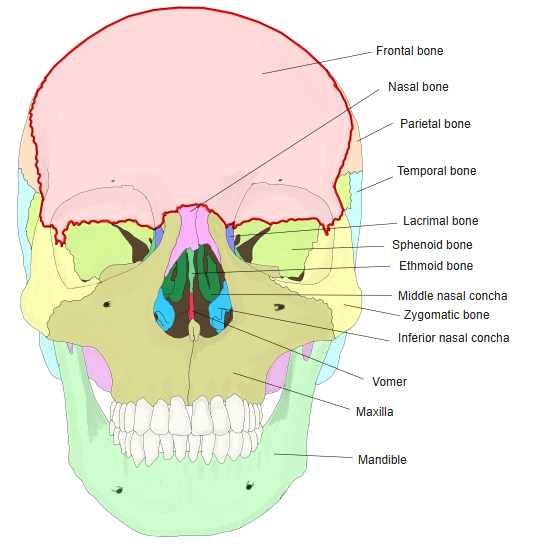
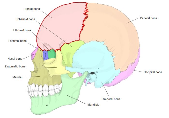
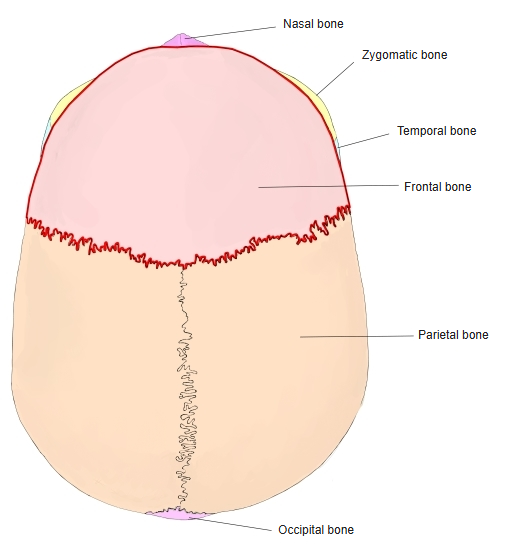
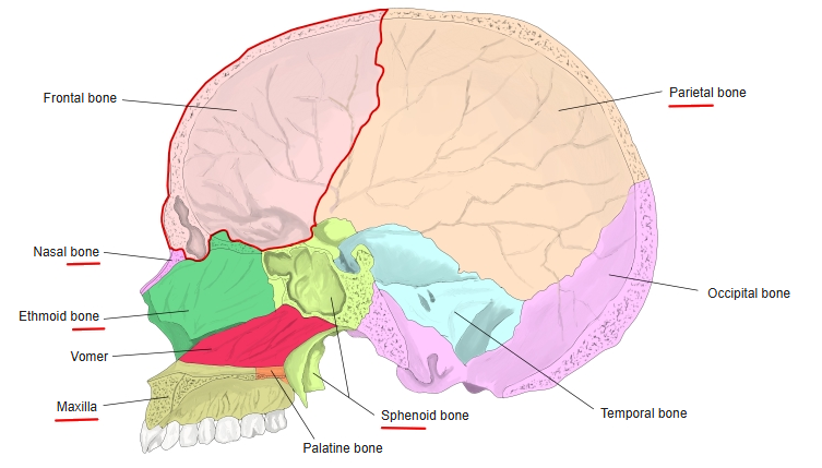
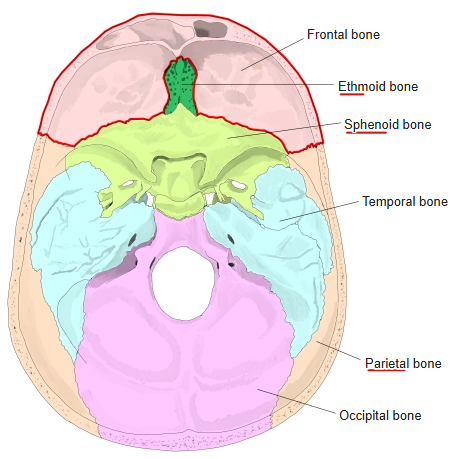
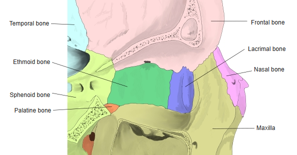
In these figures the frontal bone is outlined in red.
Muscle attachments
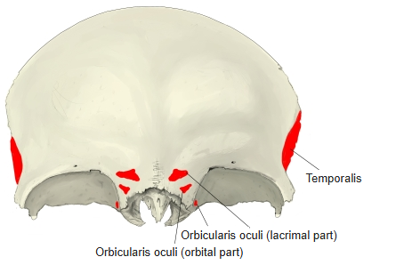
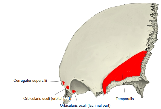
- Origin of: Temporalis, Corrugator supercilii, Orbicularis oculi
- Insertion of: none
Temporalis arises from the temporal surface below the temporal line; orbicularis oculi (orbital part) from the medial part of the orbital margin.
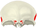
Muscle attachments (anterior) 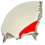
Muscle attachments (left lateral)
Borders
- Supra-orbital margin – the upper rim of the orbit.
- Medial border – surrounds the ethmoidal notch.
- Nasal border – the half-moon-shaped edge above the nasal spine, meeting the nasal bones and the frontal processes of the maxillae.
- Parietal border (posterior border) – the serrated edge meeting both parietal bones.
- Sphenoidal border – the continuation of the parietal border on the orbital part, where the greater and lesser wings of the sphenoid attach.
Foramina and openings
- Supra-orbital foramen (or notch) – supra-orbital nerve (lateral branch), supra-orbital artery and vein.
- Frontal notch (or foramen) – supra-orbital nerve (medial branch) and supratrochlear vessels.
- Anterior ethmoidal foramen – anterior ethmoidal nerve, artery and vein.
- Posterior ethmoidal foramen – posterior ethmoidal nerve, artery and vein.
- Opening of the frontal sinus – carries no nerve or vessel; the opening of the frontal sinus.
- Foramen caecum – described as transmitting an emissary vein or a process of the dura mater.
Development and variation
- The frontal bone ossifies in membrane. The main ossification centres, one on each side, appear at the frontal tubers.
- At birth the two halves are still separate; they begin to unite at about 2 years and fuse in childhood. Fusion near the root of the nose is late, and a small separate bone may remain there in the adult.
- The frontal sinus begins to form at about 2 years and is complete at about 20, though it may go on growing into old age.
- A midline suture, the metopic (frontal) suture, often persists in adults, dividing the bone in two.
- A small spine, the trochlear spine, is occasionally found beside the trochlear fovea, where the pulley of superior oblique attaches.
References and sources
- Further reading: Frontal bone, Wikipedia.