Latin: os parietale
Overview
- A paired bone of the neurocranium that forms most of the roof and sides of the cranial vault.
- It is roughly four-sided, so it has four borders and four angles, and two surfaces (external and internal).
- Each parietal bone is joined to its neighbours by sutures: the other parietal bone, the frontal, occipital, temporal and sphenoid bones.
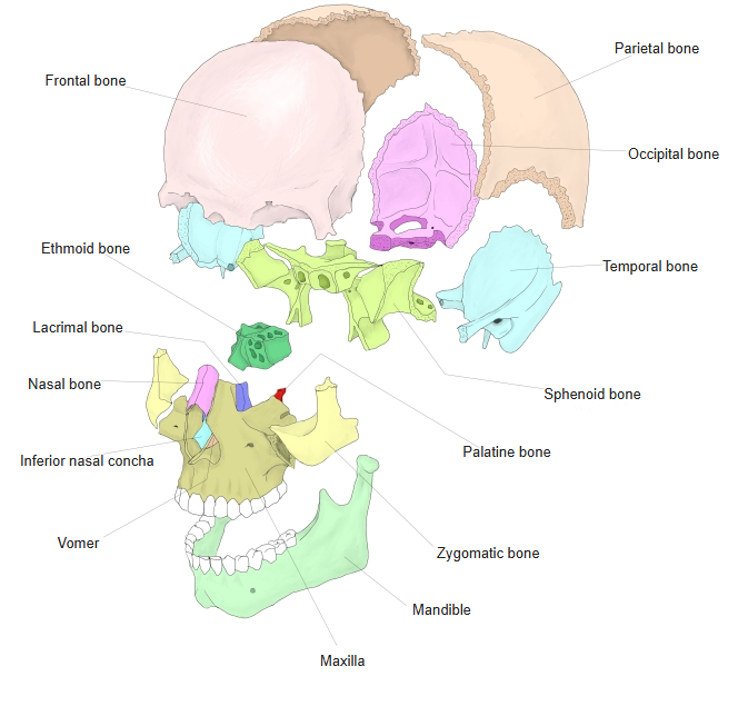
- Bones of the skull
- Neurocranium (cranial bones)
- Frontal bone
- Parietal bone (2)
- Occipital bone
- Temporal bone (2)
- Sphenoid bone
- Ethmoid bone
- Viscerocranium (facial bones)
- Nasal bone (2)
- Vomer
- Lacrimal bone (2)
- Inferior nasal concha (2)
- Maxilla (2)
- Zygomatic bone (2)
- Palatine bone (2)
- Mandible
- Hyoid bone
- Neurocranium (cranial bones)
Illustrations
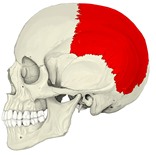
Skull (left lateral) 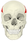
Skull (anterior) 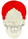
Skull (posterior) 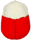
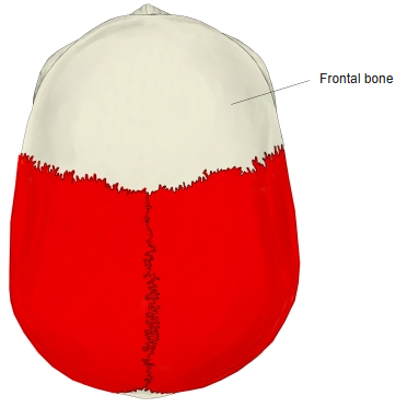
Skull (from above) 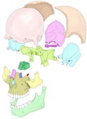
Bones of the skull (separated)
Articulations
| # | Bone | Joint |
|---|---|---|
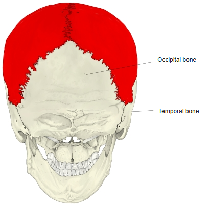
| 1 | Parietal bone of the other side | Sagittal suture |
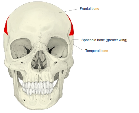
| 2 | Frontal bone | Coronal suture |
| 3 | Occipital bone | Lambdoid suture |
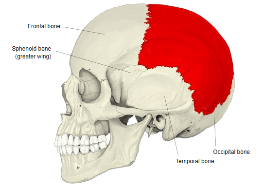
| 4 | Temporal bone | Squamous suture, parietomastoid suture |
| 5 | Sphenoid (greater wing) | Sphenoparietal suture |
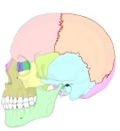
Neighbouring bones (left lateral) 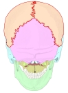
Neighbouring bones (posterior) 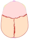
Neighbouring bones (from above) 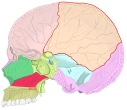
Neighbouring bones (sagittal section)
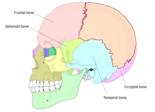
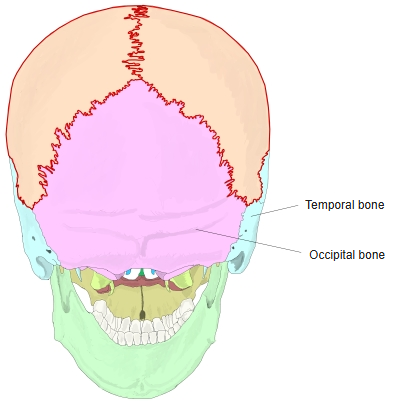
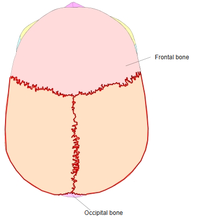
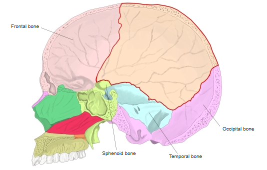
In these figures the parietal bone is outlined in red.
Muscle attachments
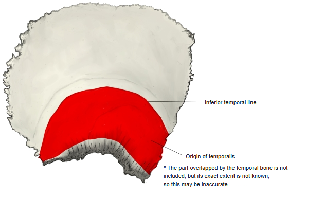
- Origin of: Temporalis
- Insertion of: none
Part of temporalis arises from the external surface below the inferior temporal line.
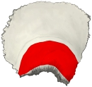
Muscle attachment (external surface)
Surfaces
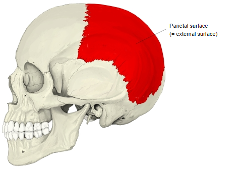
- External (parietal) surface – convex, with the parietal tuber (eminence) at its centre and the curved superior and inferior temporal lines below it.
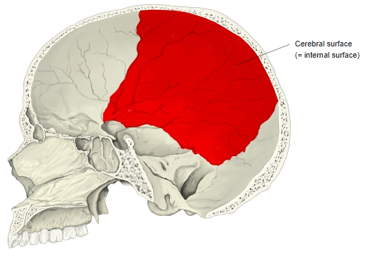
- Internal (cerebral) surface – concave, with the groove for the superior sagittal sinus along the sagittal border, grooves for the branches of the middle meningeal artery, and granular foveolae (pits for the arachnoid granulations).
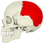
External surface 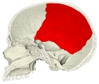
Internal surface
Borders and angles
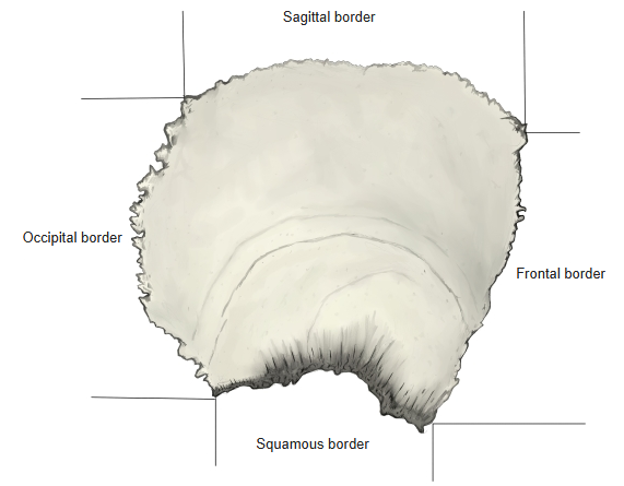
- Sagittal border – meets the other parietal bone (sagittal suture).
- Frontal border – meets the frontal bone (coronal suture).
- Occipital border – meets the occipital bone (lambdoid suture).
- Squamous border – meets the squamous part of the temporal bone (squamous suture) and the greater wing of the sphenoid.
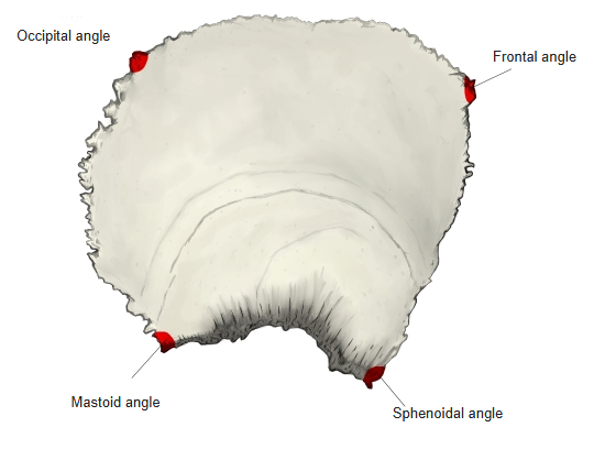
- Angles – frontal (front-upper), occipital (back-upper), sphenoidal (front-lower) and mastoid (back-lower) angles.
Borders of the right parietal bone 
Angles of the right parietal bone
Foramen
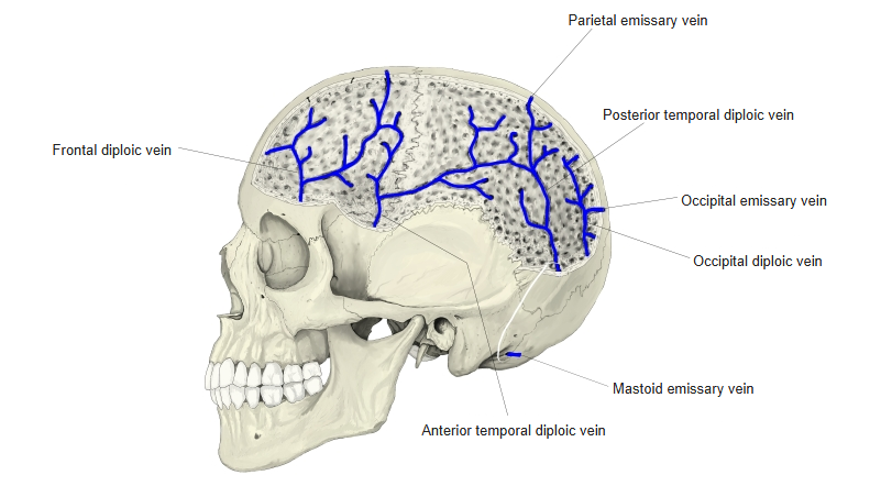
- Parietal foramen – near the sagittal border towards the back; transmits the parietal emissary vein.
Emissary and diploic veins
References and sources
- Further reading: Parietal bone, Wikipedia.