Latin: Vena brachiocephalica
Overview
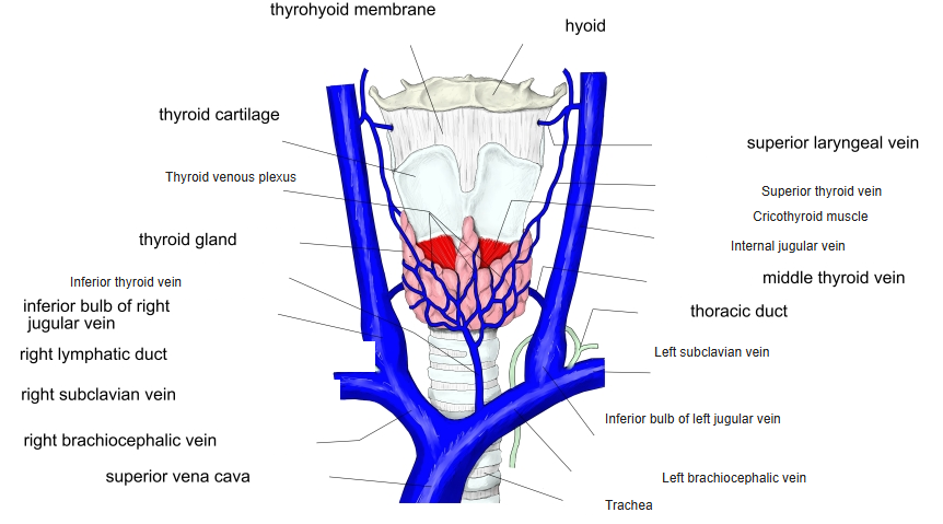
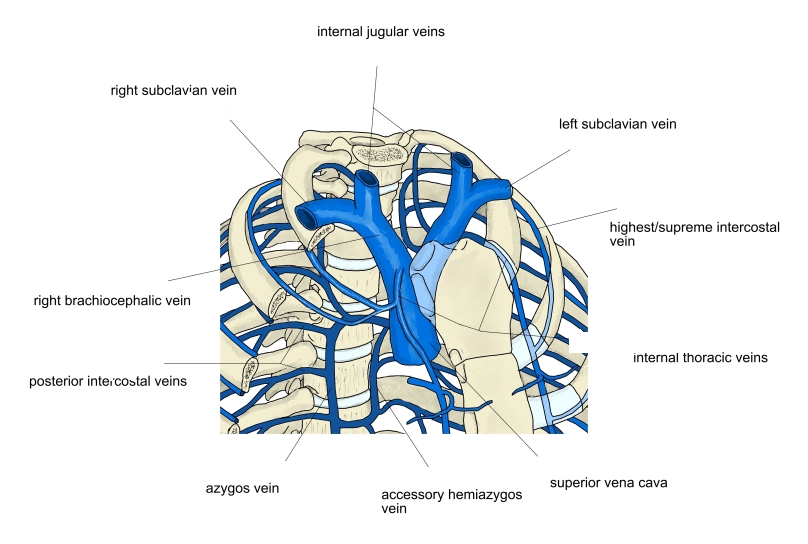
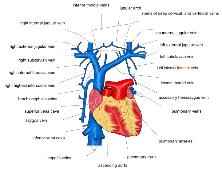
- Two large veins, right and left, formed behind the sternoclavicular joints by the union of the internal jugular and subclavian veins.
- The right vein is short (about 2.5 cm) and runs almost vertically down.
- The left vein is long (about 6 cm) and crosses obliquely to the right behind the manubrium, in front of the branches of the aortic arch.
- The two unite behind the first right costal cartilage to form the superior vena cava. They have no valves.
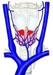
Veins of the thyroid gland and larynx 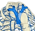
Formation of the superior vena cava
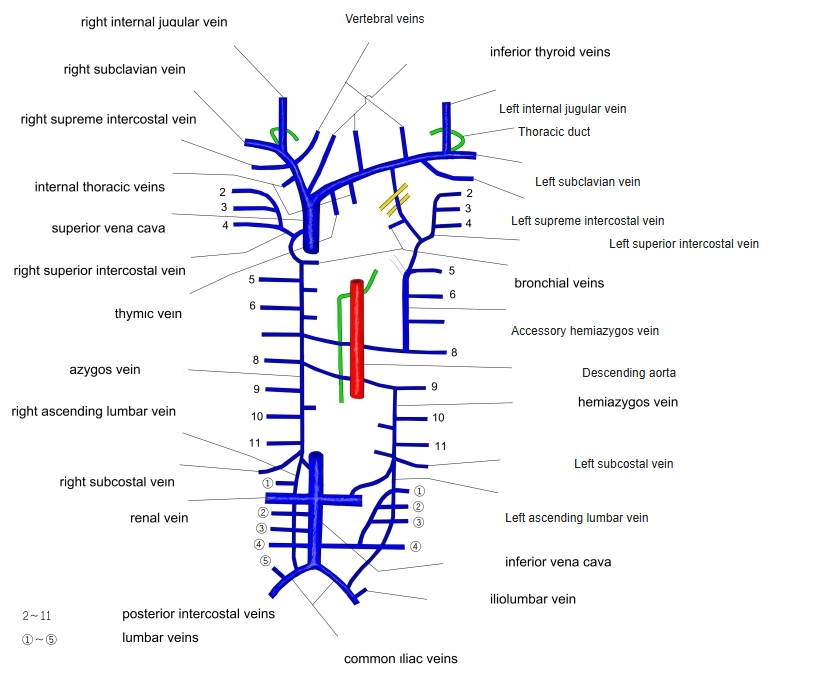
Tributaries
| Side | Tributaries |
|---|---|
| Both | Vertebral vein, internal thoracic vein, inferior thyroid veins, first posterior intercostal vein |
| Left only | Left superior intercostal vein, thymic and pericardiac veins |
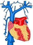
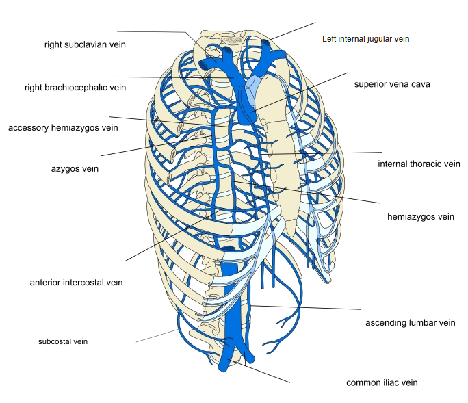
Superior vena cava and its tributaries 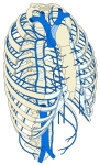
Veins of the thoracic wall 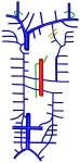
Veins of the thorax (diagram)
Lymphatic ducts
- The thoracic duct opens at the junction of the left internal jugular and subclavian veins (the left venous angle); the right lymphatic duct opens at the right venous angle.
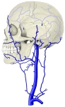
Veins of the head and neck (side view) 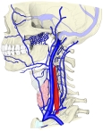
Tributaries of the jugular veins 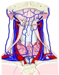
Veins of the front of the neck
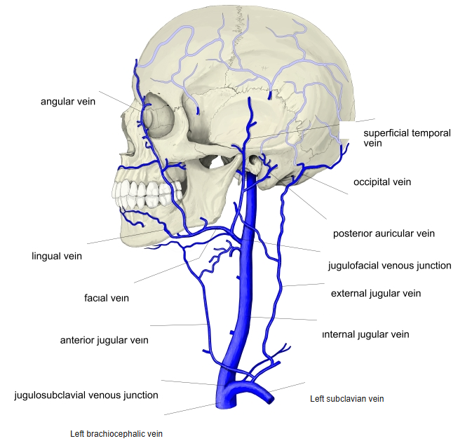
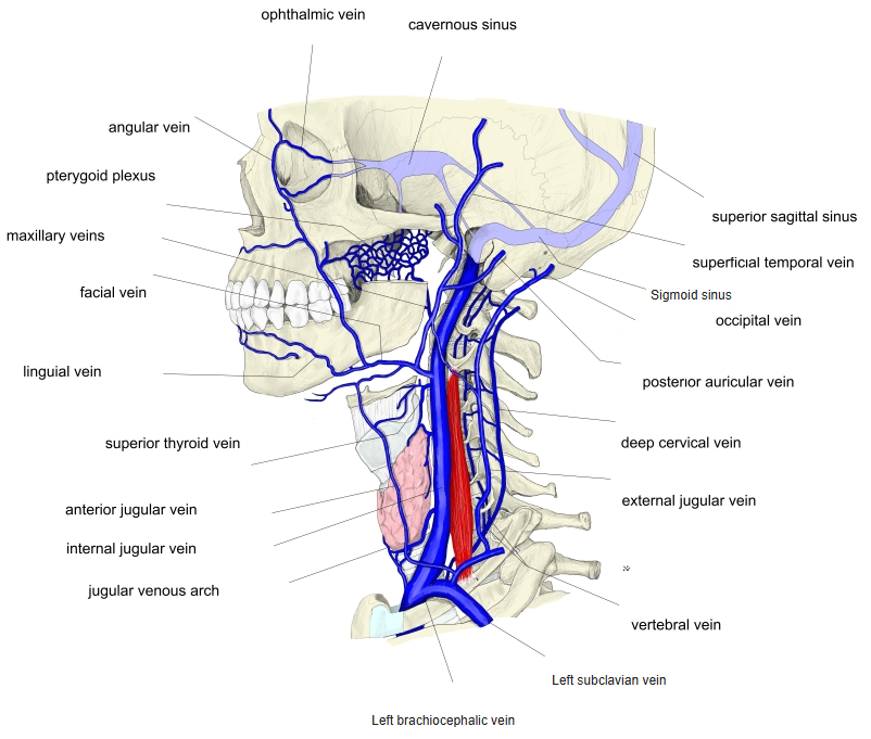
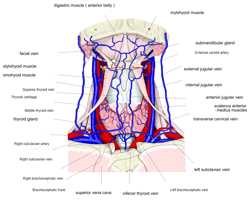
Clinical notes
- The left vein can be compressed between the sternum and the aortic arch.
- Central venous catheters pass through it to reach the superior vena cava.
References and sources
- Further reading: Brachiocephalic vein, Wikipedia.