Latin: Vena cava superior
Overview
- The large vein that returns blood from the head, neck, upper limbs and chest wall to the heart.
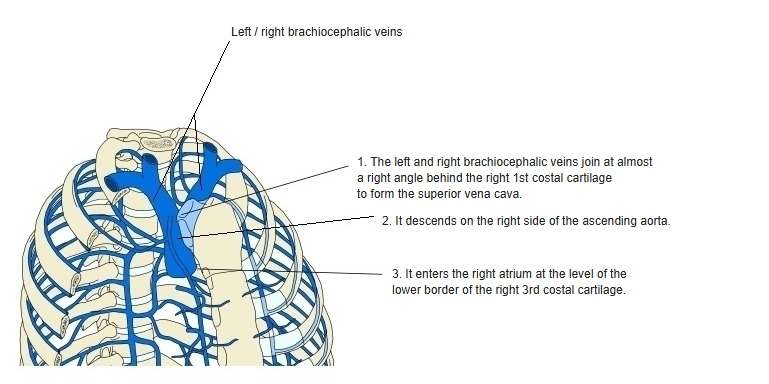
- Formed by the union of the right and left brachiocephalic veins behind the lower border of the right 1st costal cartilage.
- Short (about 7 cm) and without valves; it ends in the upper part of the right atrium.
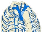
Course of the superior vena cava 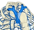
Tributaries of the superior vena cava
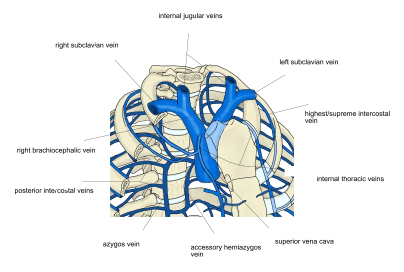
Course
- Descends vertically on the right side of the ascending aorta, behind the right border of the sternum.
- Its lower half lies within the fibrous pericardium.
- Enters the right atrium at the level of the right 3rd costal cartilage.
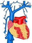
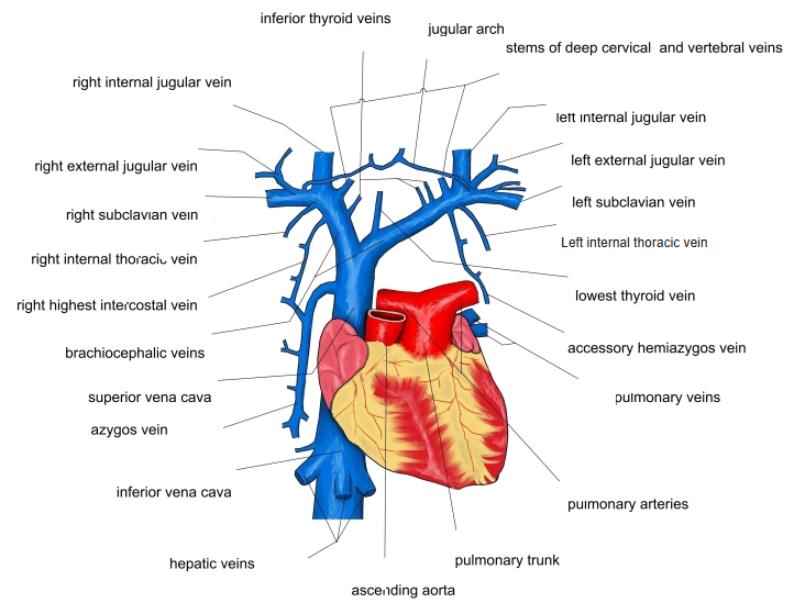
Veins entering the superior vena cava 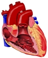
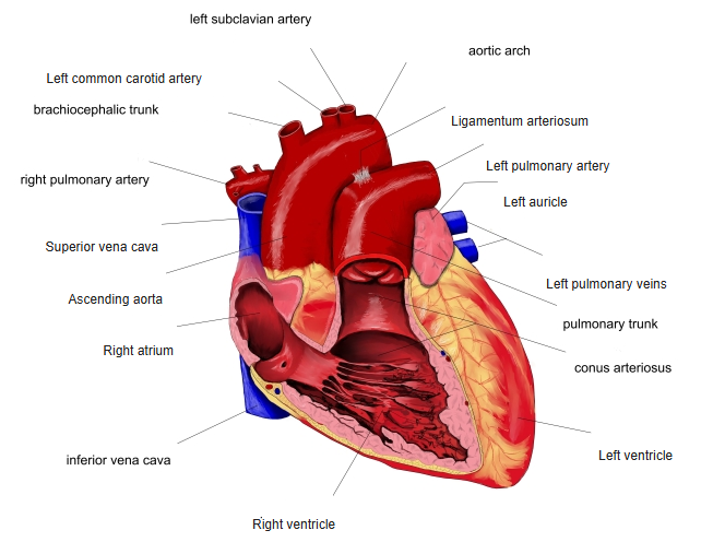
Heart and great vessels (front view)
Tributaries
| Vein | Drains |
|---|---|
| Right and left brachiocephalic veins | Head and neck (internal jugular veins) and upper limbs (subclavian veins) |
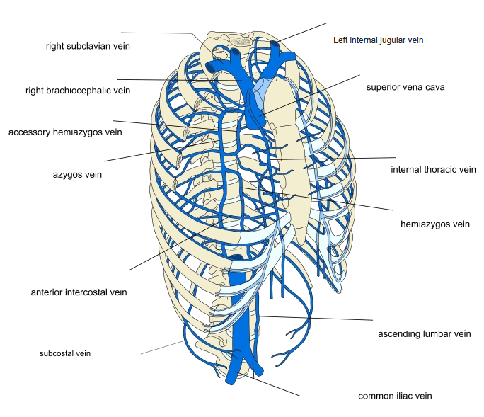
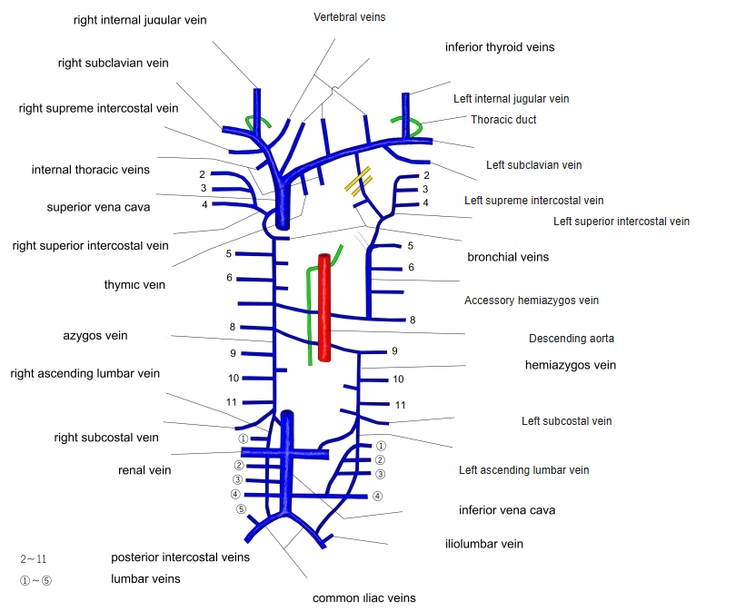
| Azygos vein | Posterior thoracic wall; arches over the right lung root and enters from behind at the level of T4 |
| Small pericardial and mediastinal veins | Pericardium and mediastinum |
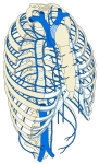
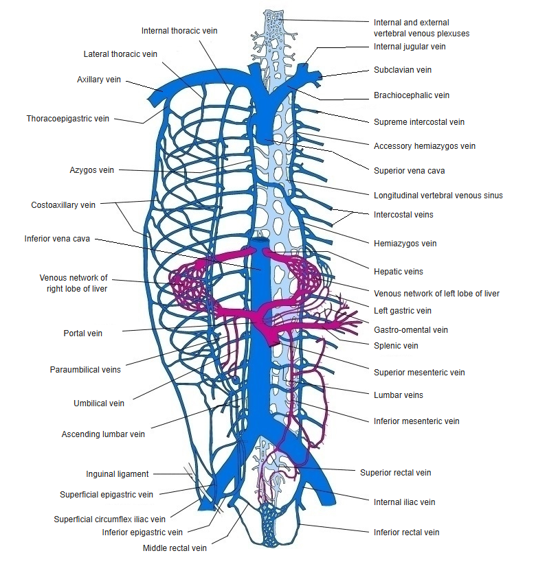
Superior vena cava and the azygos system
Relations
- In front: the right lung and pleura, and the thymus (or its remnant).
- Behind: the root of the right lung and the right vagus nerve.
- On the right: the right phrenic nerve and the pleura; on the left: the ascending aorta.
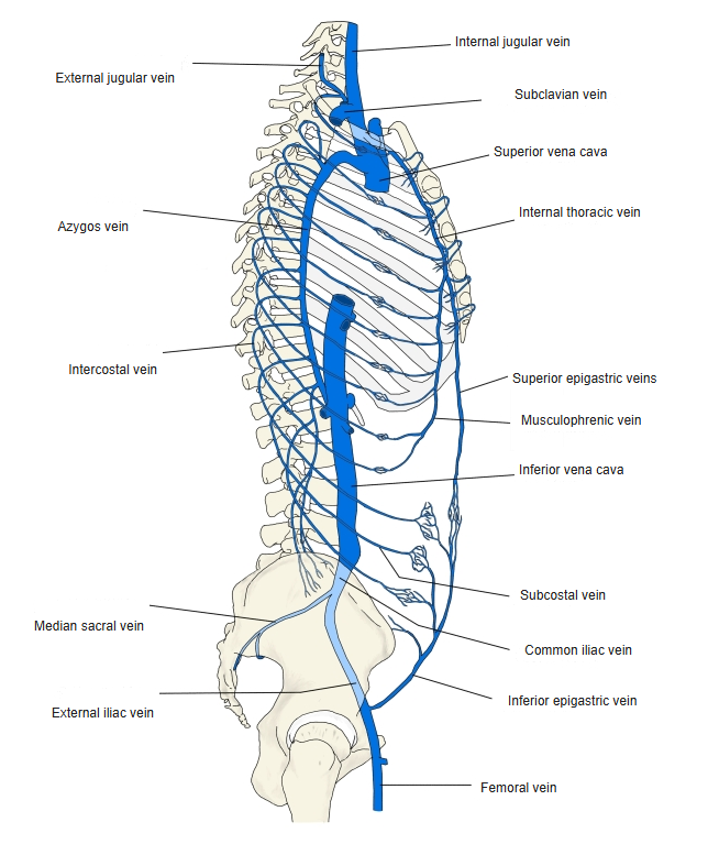
Veins of the trunk (side view) 
Main veins of the trunk
Clinical notes
- Superior vena cava syndrome: obstruction (most often by a lung tumour or enlarged lymph nodes) causes swelling of the face, neck and arms and distended neck veins.
- Central venous catheters are usually placed with their tip in the lower superior vena cava.
References and sources
- Further reading: Superior vena cava, Wikipedia.