Latin: Vena femoralis
Overview
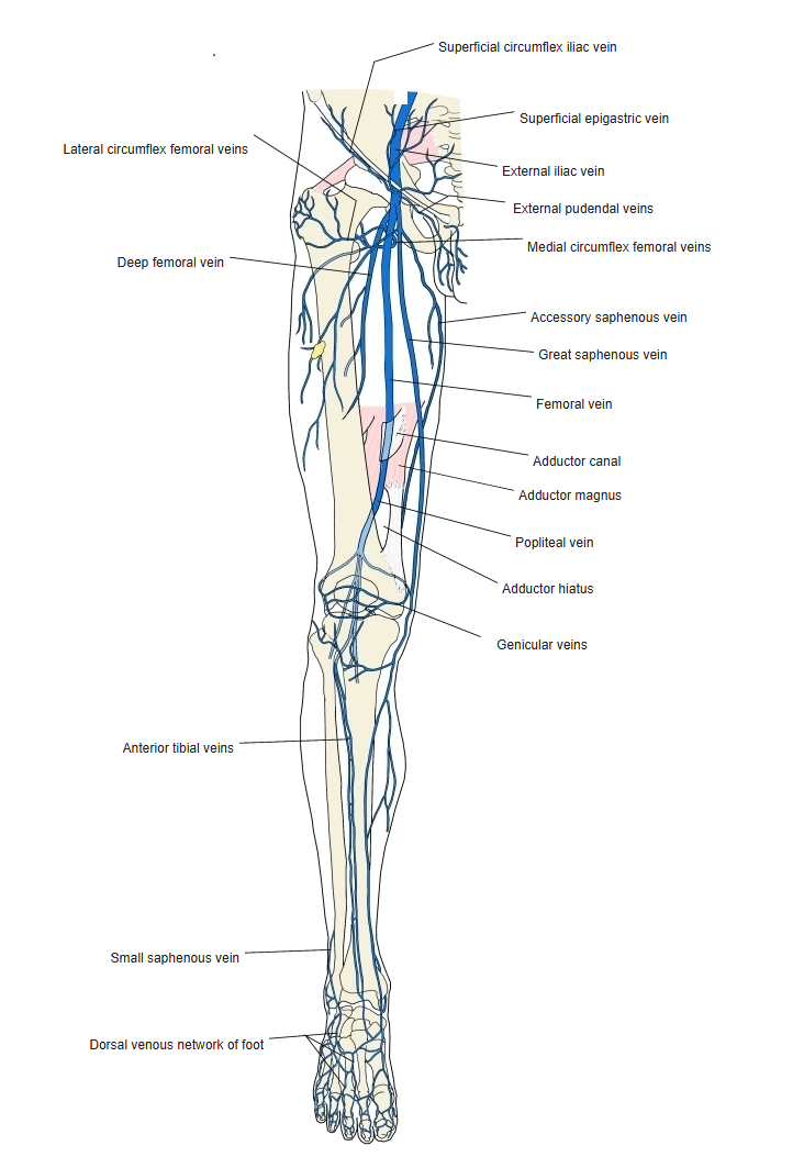
- The main deep vein of the thigh; it accompanies the femoral artery.
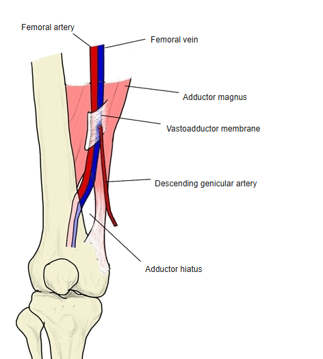
- Begins at the adductor hiatus as the continuation of the popliteal vein.
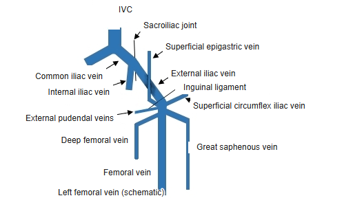
- Ends behind the inguinal ligament, where it becomes the external iliac vein.
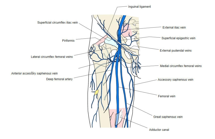
Veins of the lower limb (front view)
Course
- Passes up through the adductor canal, first behind and then medial to the femoral artery.
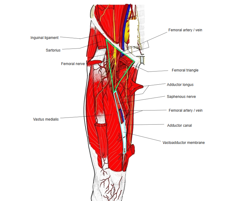
- In the femoral triangle it lies medial to the artery, inside the femoral sheath; the femoral nerve lies lateral to the artery.
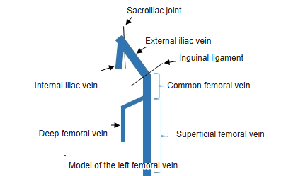
- The part above the deep femoral vein is called the common femoral vein and the part below it the superficial femoral vein in clinical use.
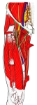
Femoral triangle 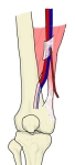
Adductor canal and adductor hiatus
Tributaries
- Deep femoral vein.
- Great saphenous vein (at the saphenous opening).
- Lateral and medial circumflex femoral veins, muscular veins.
Veins of the front of the thigh
Clinical notes
- Lies just medial to the femoral pulse below the inguinal ligament: a site for central venous access.
- A common site of deep vein thrombosis.
References and sources
- Further reading: Femoral vein, Wikipedia.