Femoral triangle (Latin: trigonum femorale)
Also called Scarpa's triangle
· Also called Scarpa's triangle.
· Below, it continues into the adductor canal.
Other structures found there include:
· Lateral femoral cutaneous nerve: a branch of the lumbar plexus from the second and third lumbar nerves; a sensory nerve.
· Nerve to pectineus: one of the muscular branches of the femoral nerve.
· Femoral branch of the genitofemoral nerve (inside the femoral sheath).
· Deep inguinal lymph nodes (inside the femoral sheath).
In the iliopectineal fossa lie the femoral nerve and the femoral artery and vein.
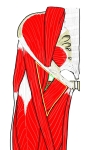 Femoral triangle and iliopectineal fossa
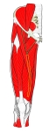 Femoral triangle and adductor canal
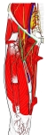 Vessels and nerves of the thigh
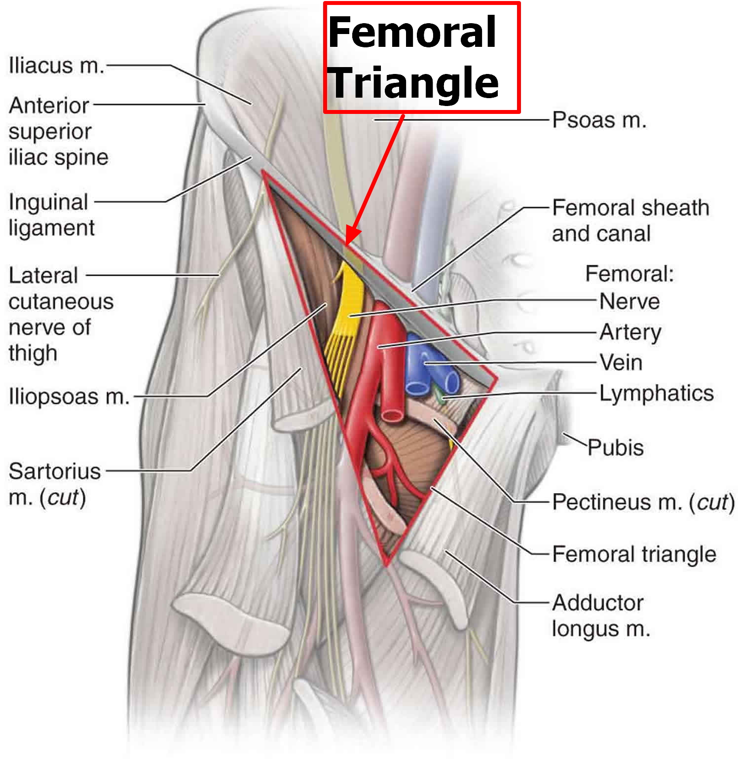
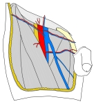 Contents of the triangle (diagram)
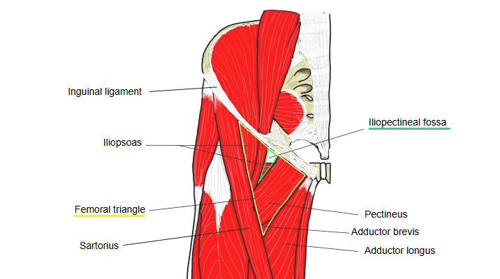
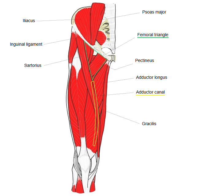
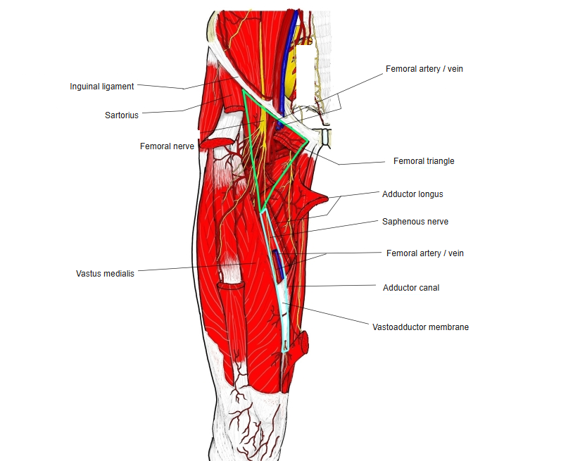
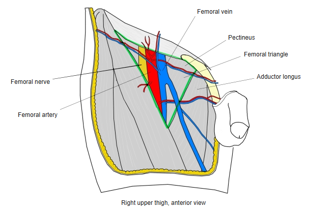
A standard description:
The femoral triangle is the region in the upper front of the thigh bounded by the inguinal ligament, the medial border of sartorius and the medial border of adductor longus (some texts take its lateral border, which gives a smaller triangle); below, it continues into the adductor canal. Its deep upper medial part is the iliopectineal fossa between pectineus and iliopsoas, which holds the femoral artery and vein, lymph nodes and lymph vessels. It is seen as a hollow below the groin when the thigh is flexed, abducted and laterally rotated; the femoral pulse is felt here, and the femoral vessels are a common route for catheters.
Reference websites
[Wikipedia]
· Femoral triangle, Wikipedia.
· Femoral sheath, Wikipedia.
[Websites with illustrations]
· Femoral triangle with the femoral nerve, artery, vein and lymphatics (Health Jade)
· Femoral triangle and adductor canal (SlideShare)

· Femoral triangle: nerves and vessels below the inguinal ligament (WordPress)

Boundaries and contents
The boundaries of the femoral triangle:
· Base (superior): the inguinal ligament.
· Medial: the medial border of adductor longus (some texts use its lateral border, which makes the triangle smaller).
· Lateral: the medial border of sartorius.
Apex: where sartorius crosses adductor longus; it continues into the adductor canal. Roof: skin, superficial fascia and the fascia lata, which has the saphenous opening in it, covered by the cribriform fascia; the superficial fascia holds the superficial inguinal lymph nodes, the femoral branch of the genitofemoral nerve and the upper part of the great saphenous vein. Floor: iliopsoas laterally and pectineus and adductor longus medially.
Many important structures pass through the triangle just under the skin. From lateral to medial they are the femoral nerve, the femoral artery, the femoral vein and the deep inguinal lymph nodes (often remembered as "NAVeL"):
Lateral femoral cutaneous nerve: crosses near the lateral angle of the triangle on its way to the skin of the side of the thigh.
Femoral nerve and its branches: it enters under the inguinal ligament in the groove between iliacus and psoas major, lateral to the femoral artery and outside the femoral sheath, and soon divides into its branches.
Nerve to pectineus: runs behind the femoral sheath to the front of pectineus.
The femoral sheath encloses the upper few centimetres of the femoral artery and vein; the nerve lies outside it. It has three compartments, from lateral to medial:
Lateral compartment: the femoral artery and the femoral branch of the genitofemoral nerve, which supplies the skin over the triangle.
Femoral artery: enters at the base of the triangle at the mid-inguinal point and leaves through the apex into the adductor canal; the deep femoral artery arises from it in the triangle.
Intermediate compartment: the femoral vein, medial to the artery at the base and behind it towards the apex. The great saphenous vein passes through the saphenous opening to join it in the triangle.
Medial compartment (the femoral canal): lymph vessels and deep inguinal lymph nodes, which drain the deep lymph vessels of the lower limb and the superficial inguinal nodes.
Where it is found
Related terms
- Saphenous opening
- Adductor canal
- Femoral sheath
- Inguinal ligament
- Femoral artery
- Femoral vein
- Femoral nerve
References and sources
- Further reading: Femoral triangle, Wikipedia.
- Further reading: Femoral sheath, Wikipedia.
- Federative International Programme for Anatomical Terminology (FIPAT). Terminologia Anatomica, 2nd ed. (TA2), 2019.