Latin: Arteria thoracica interna
Overview
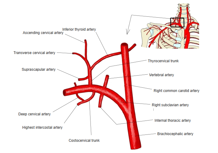
- A branch of the 1st part of the subclavian artery that supplies the front of the thoracic wall and the breast; also called the internal mammary artery.
- Descends behind the upper six costal cartilages, about 1 cm lateral to the sternum.
- Ends in the 6th intercostal space by dividing into the musculophrenic artery and the superior epigastric artery.
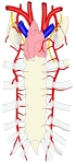
Internal thoracic arteries 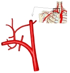
Branches of the subclavian artery (right side)
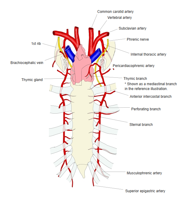
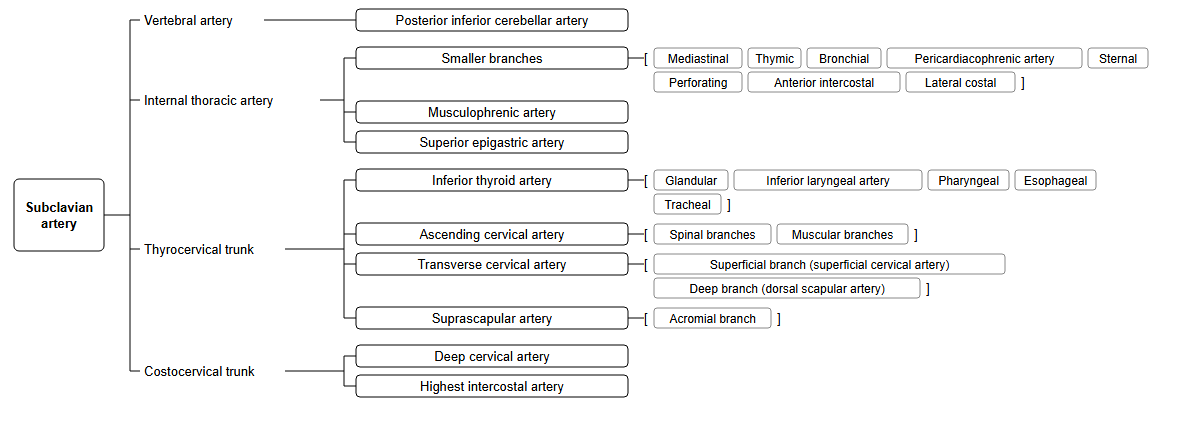
Branches
| Branch | Supply |
|---|---|
| Pericardiacophrenic artery | Pericardium and diaphragm, with the phrenic nerve |
| Mediastinal and thymic branches | Mediastinum and thymus |
| Sternal branches | Sternum and the transversus thoracis |
| Anterior intercostal branches | Upper six intercostal spaces; join the posterior intercostal arteries |
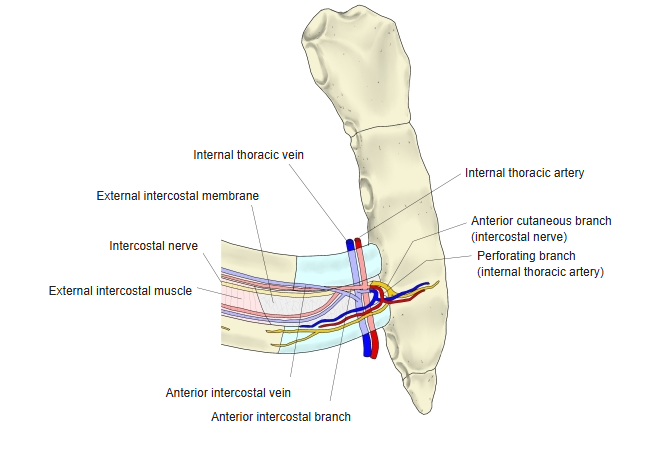
| Perforating branches | Pectoralis major and skin; the 2nd–4th give mammary branches to the breast |
| Musculophrenic artery | Diaphragm and the lower intercostal spaces |
| Superior epigastric artery | Rectus abdominis; joins the inferior epigastric artery |
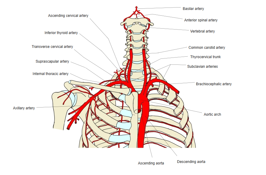
Subclavian arteries and their branches
Intercostal arteries
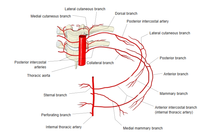
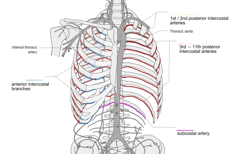
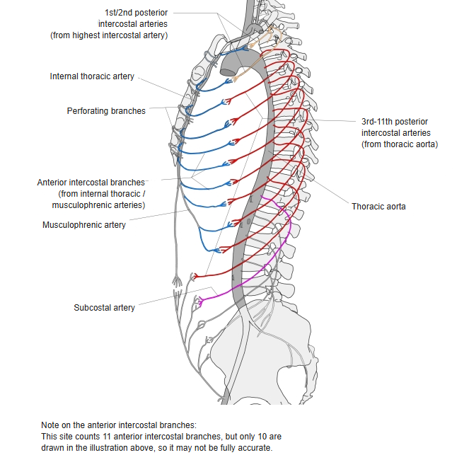
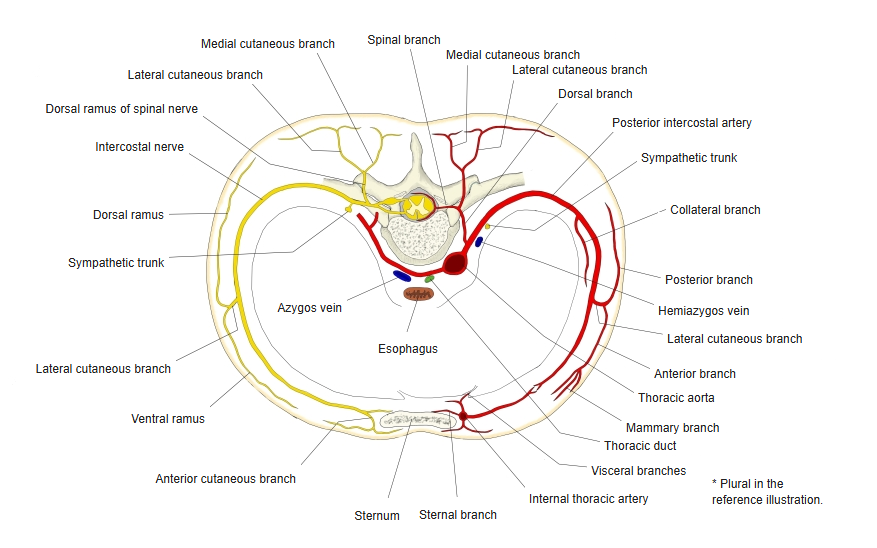
- Each intercostal space receives a posterior intercostal artery from the thoracic aorta (the 1st and 2nd from the highest intercostal artery).
- In front it is joined by anterior intercostal branches from the internal thoracic artery or the musculophrenic artery.
Intercostal arteries in one intercostal space 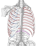
Intercostal arteries of the thorax 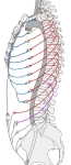
Anterior and posterior intercostal arteries (side view) 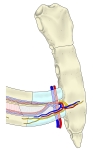
Perforating branch of the internal thoracic artery 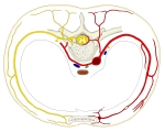
Intercostal artery and nerve (cross section)
Clinical notes
- The internal thoracic artery is the most commonly used vessel for coronary artery bypass grafting.
References and sources
- Further reading: Internal thoracic artery, Wikipedia.