Latin: Arteriae intercostales posteriores
Overview
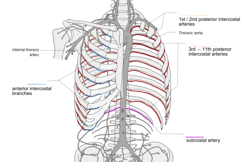
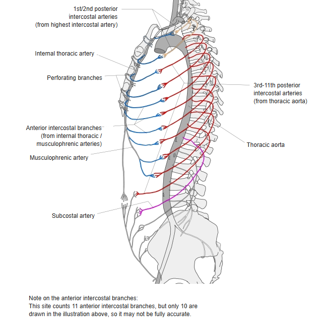
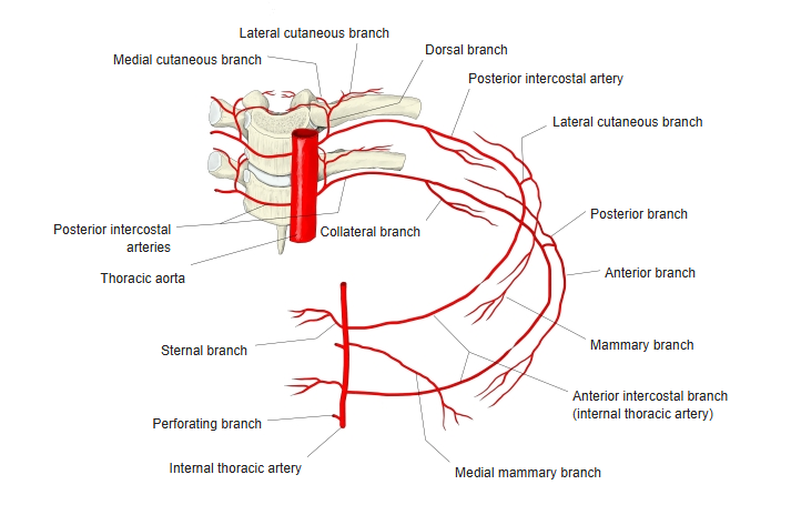
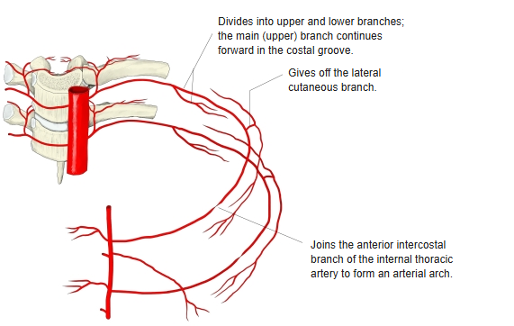
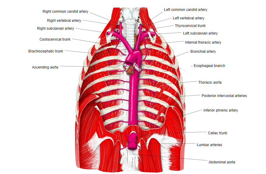
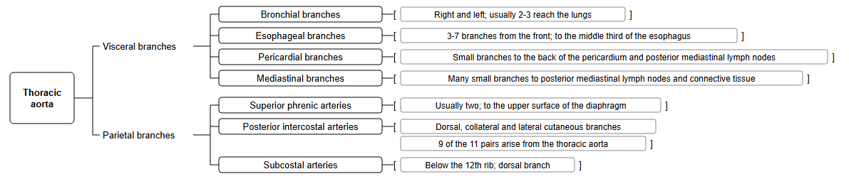
- Paired arteries that run in the intercostal spaces. The 1st and 2nd come from the highest (supreme) intercostal artery of the costocervical trunk; the 3rd–11th come from the thoracic aorta.
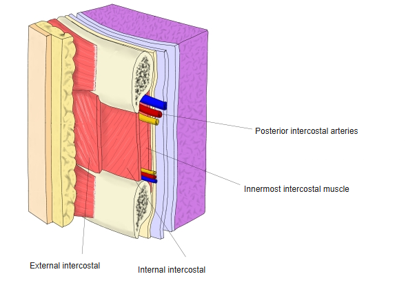
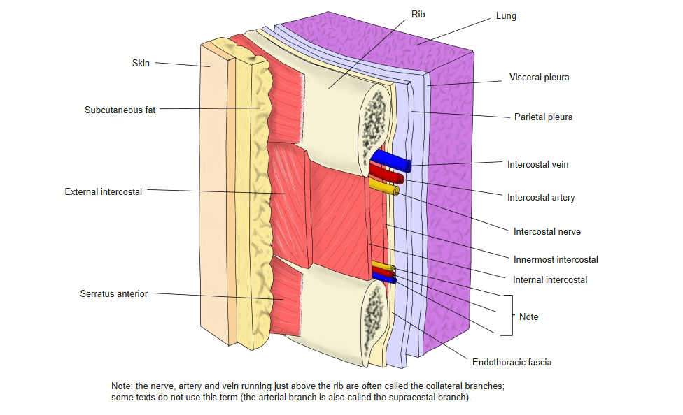
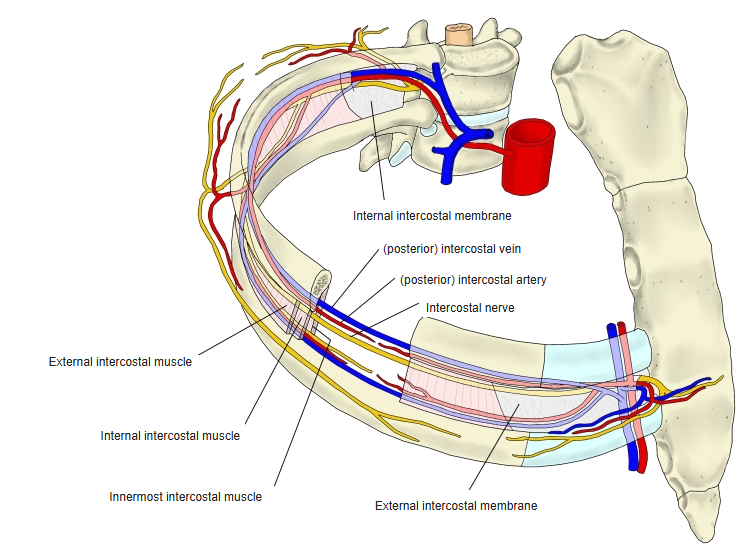
- Each runs in the costal groove on the lower border of the rib above, between the intercostal vein (above) and the intercostal nerve (below).
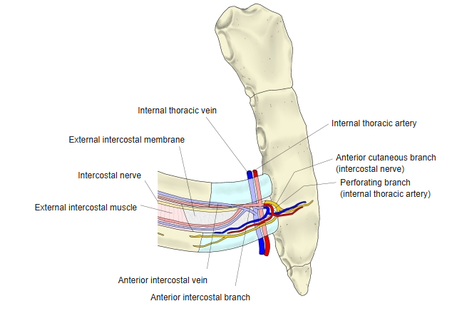
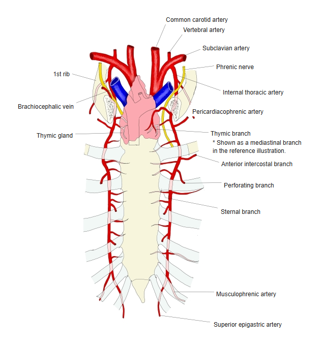
- Joins the anterior intercostal branches of the internal thoracic artery in front.
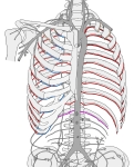
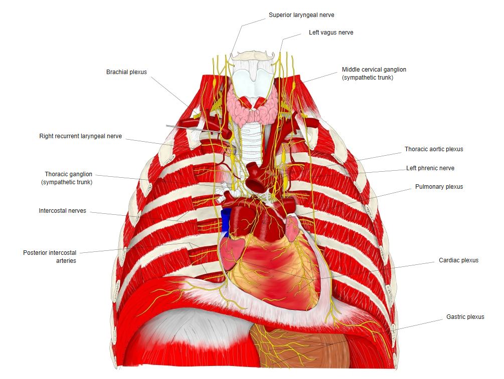
Intercostal arteries of the thorax 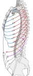
Anterior and posterior intercostal arteries (side view) 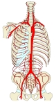
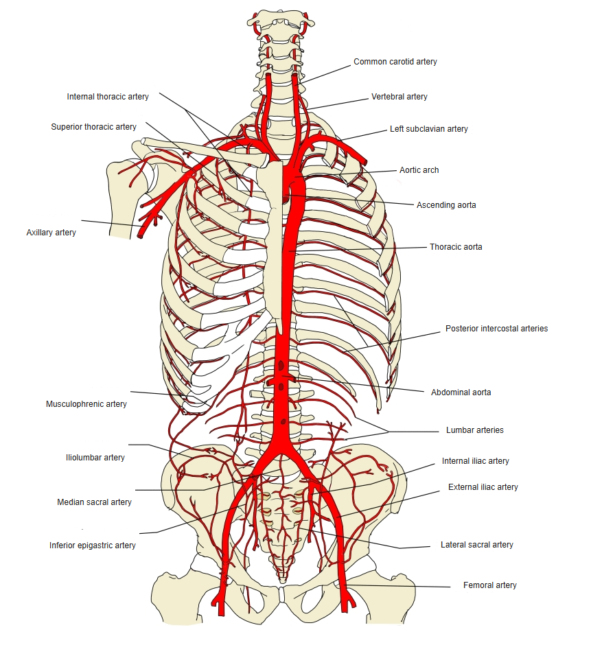
Aorta and its branches in the trunk (front view) 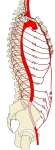
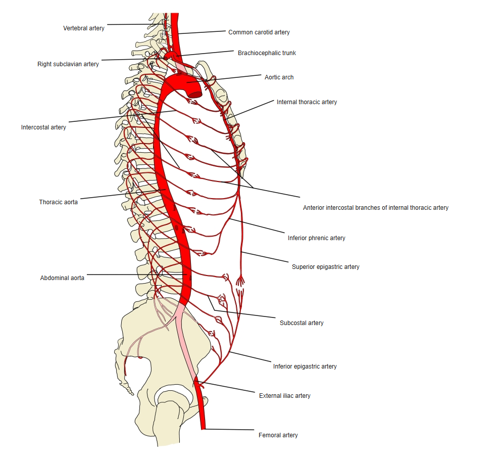
Aorta and its branches in the trunk (side view)
Branches
| Branch | Supply |
|---|---|
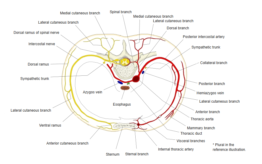
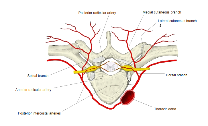
| Dorsal branch | Back muscles and skin; gives a spinal branch to the vertebral canal and spinal cord |
| Collateral branch | Runs along the upper border of the rib below |
| Lateral cutaneous branch | Skin of the side of the thorax; lateral mammary branches |
| Muscular branches | Intercostal muscles |
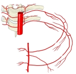
Intercostal arteries in one intercostal space 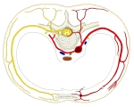
Intercostal artery and nerve (cross section) 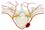
Branches of a posterior intercostal artery 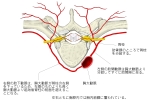
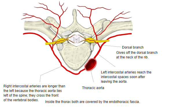
Origin of the posterior intercostal arteries 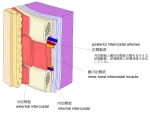
Posterior intercostal artery and nerve 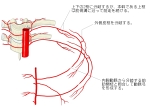
Course of a posterior intercostal artery 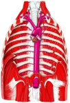
Thoracic aorta and its branches
In the intercostal space
- From above down in the costal groove: vein, artery, nerve (VAN).
- Needles placed into the chest wall are passed just above the upper border of a rib to avoid these vessels.
Intercostal vessels and nerves 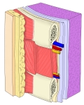
Intercostal nerve and its branches 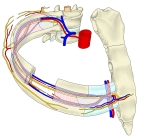
Intercostal space 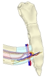
Perforating branch of the internal thoracic artery 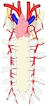
Internal thoracic arteries
References and sources
- Further reading: Posterior intercostal arteries, Wikipedia.