Latin: Cavitas pericardiaca
Overview
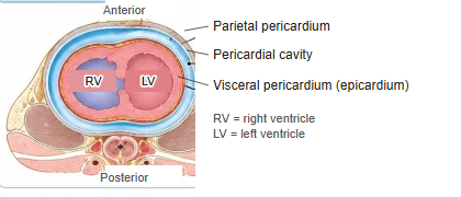
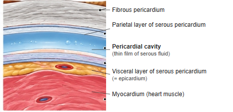
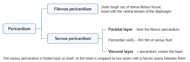
- The narrow potential space between the parietal and visceral layers of the serous pericardium.
- Normally holds only about 15–50 mL of serous fluid, which lets the heart beat with little friction.
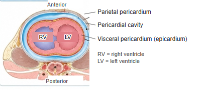
Pericardium in a cross section of the thorax 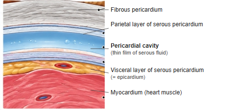
Layers of the pericardium (enlarged section)
Pericardial sinuses
- Transverse pericardial sinus: between the arterial outflow (aorta and pulmonary trunk) in front and the veins behind.
- Oblique pericardial sinus: a recess behind the left atrium, bounded by the pulmonary veins and inferior vena cava.
Clinical notes
- Pericardial effusion: too much fluid in the cavity; if it gathers quickly it causes cardiac tamponade.
- Pericardiocentesis: fluid is drawn off with a needle, usually from below the xiphoid process.
References and sources
- Further reading: Pericardial cavity, Wikipedia.