Latin: Pericardium
Overview
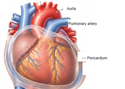
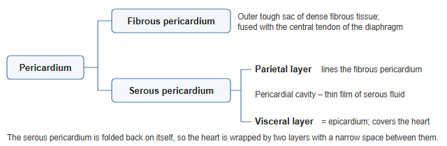
- The double-walled sac that encloses the heart and the roots of the great vessels.
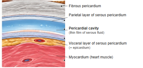
- Two parts: the outer fibrous pericardium and the inner serous pericardium.
- The serous pericardium has a parietal layer and a visceral layer (epicardium) with the pericardial cavity between them.
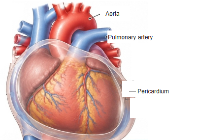
Heart inside the pericardium (front view) 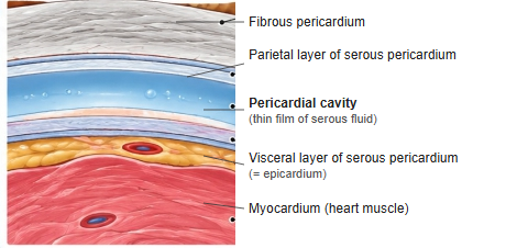
Layers of the pericardium (enlarged section)
Relations
- Below: fused with the central tendon of the diaphragm.
- In front: the sternum and costal cartilages (attached by sternopericardial ligaments).
- Behind: the oesophagus and descending aorta. On each side: the mediastinal pleura, with the phrenic nerve between.
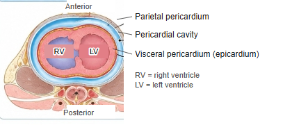
Pericardium in a cross section of the thorax
Pericardial sinuses
- Transverse pericardial sinus: a passage behind the aorta and pulmonary trunk and in front of the superior vena cava.
- Oblique pericardial sinus: a blind recess behind the left atrium.
Blood supply and innervation
- Arteries: mainly the pericardiacophrenic arteries (from the internal thoracic arteries).
- Nerves: the fibrous and parietal layers are supplied by the phrenic nerves (pain felt in the shoulder region); the visceral layer is insensitive to pain.
Clinical notes
- Pericarditis: inflammation, with sharp chest pain and a pericardial friction rub.
- Cardiac tamponade: fluid or blood in the pericardial cavity compresses the heart and reduces its filling.
References and sources
- Further reading: Pericardium, Wikipedia.