Latin: Arteriae ciliares posteriores breves
Overview
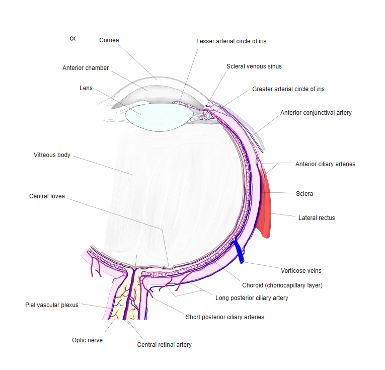
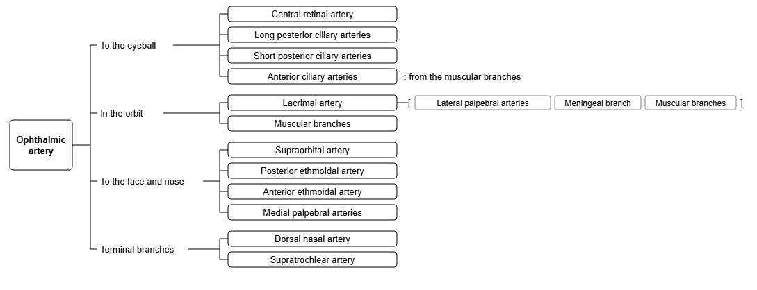
- Six to twelve branches of the ophthalmic artery.
- Pierce the sclera around the entry of the optic nerve.
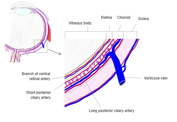
- Supply the choroid (and through it the outer layers of the retina) and the optic disc.
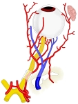
Arteries and veins of the orbit 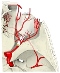
Ophthalmic artery and its branches
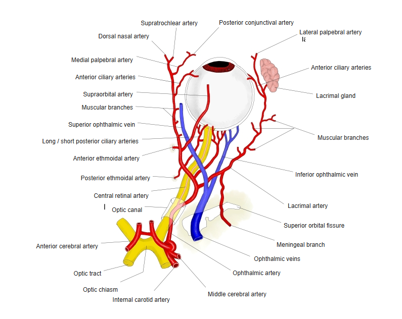
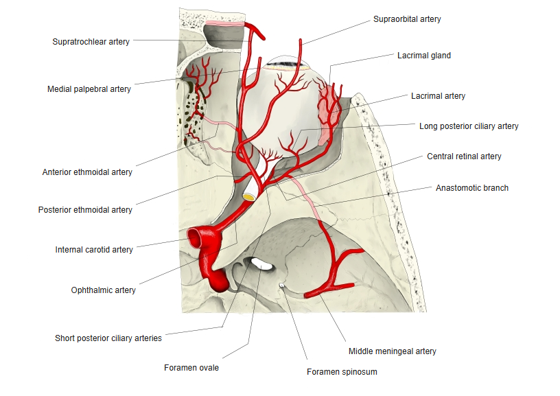
Illustrations
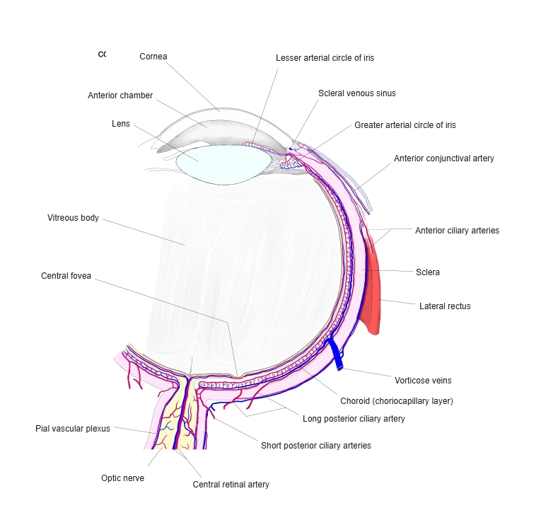
Blood supply of the eyeball 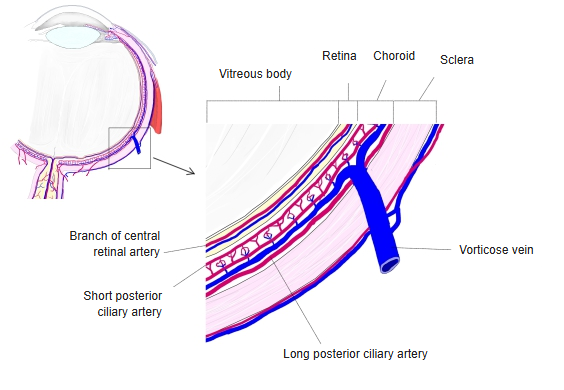
Arteries of the choroid 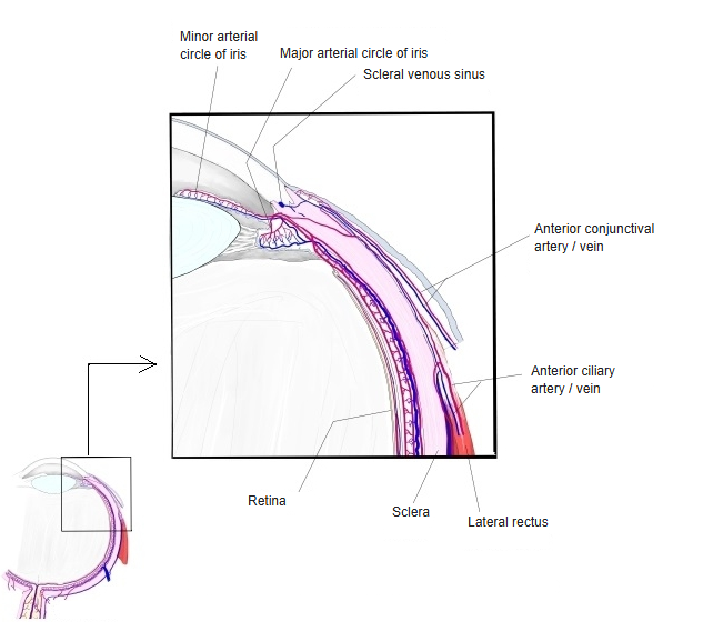
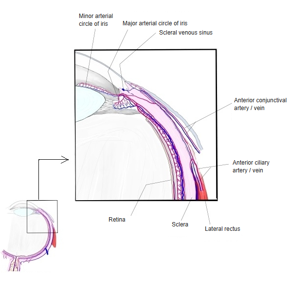
Arteries near the corneal margin 
Branches of the ophthalmic artery (chart) 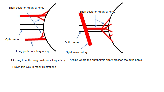
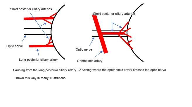
Origin of the short posterior ciliary arteries (diagram)
Clinical notes
- Their blockage is the usual cause of anterior ischaemic optic neuropathy, sudden painless loss of vision in one eye.
References and sources
- Further reading: Short posterior ciliary arteries, Wikipedia.