Latin: Sinus sigmoideus
Overview
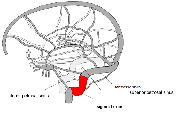
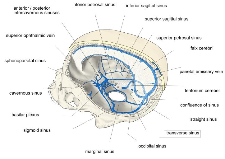
- S-shaped continuation of the transverse sinus.
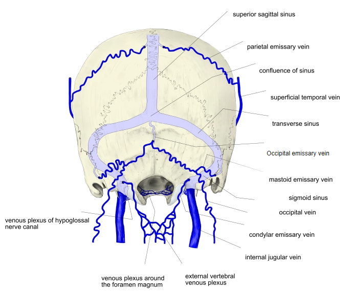
- Runs in a deep groove on the mastoid part of the temporal bone, then curves forward to the jugular foramen.
- Continues as the internal jugular vein (at the superior bulb).
Sigmoid sinus 
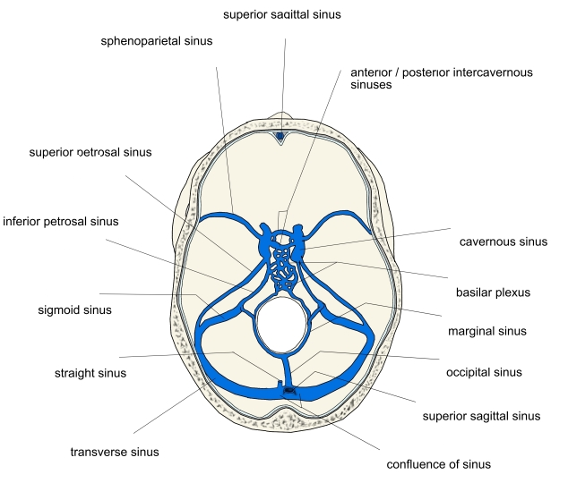
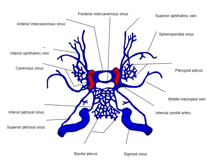
Dural venous sinuses of the cranial base
Tributaries and connections
- Mastoid and condylar emissary veins connect it with veins outside the skull.
- Small cerebellar and tympanic veins.
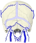
Emissary veins and dural sinuses (back view) 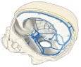
Dural venous sinuses (side view) 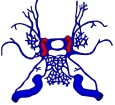
Sinuses around the cavernous sinus
Clinical notes
- Only a thin plate of bone separates it from the mastoid air cells: middle ear and mastoid infection can cause sigmoid sinus thrombosis.
References and sources
- Further reading: Sigmoid sinus, Wikipedia.