Mastoid process (Latin: processus mastoideus)
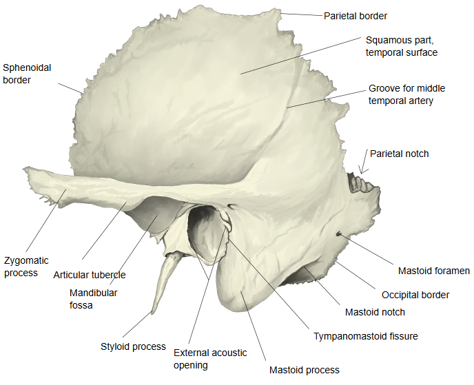
· Medial to the notch lies the groove for the occipital artery, which carries the occipital artery.
- Squamous part : occupies the middle and the whole upper part of the bone; it has two surfaces, temporal and cerebral.
- Petrous part
- Mastoid part : below the squamous part, from the mastoid process up to the parietal notch.Related terms: mastoid process, mastoid notch, groove for occipital artery, groove for sigmoid sinus, mastoid foramen, occipital border, stylomastoid foramen, canaliculus for chorda tympani, petrotympanic fissure
- Pyramid : a three-sided pyramid running forwards and medially from the boundary of the squamous and mastoid parts, with 3 surfaces and 3 borders: the petrous part in the narrow sense.
- Tympanic part : the smallest part: a thin, half-tube-shaped plate that forms the front and lower walls of the external acoustic meatus.
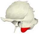 Mastoid process (lateral view)
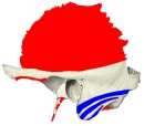 Muscle attachments (lateral view)
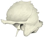 Temporal bone (lateral view), labelled
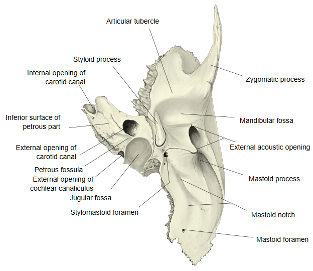
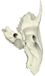 Temporal bone (inferior view), labelled
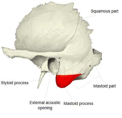
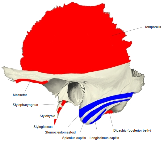
Most of the mastoid part is taken up by the large mastoid process, which projects downwards and forwards. Its surface is rough where the sternocleidomastoid is inserted. In adults the inside of the process is mostly filled with many small mastoid air cells. They enlarge after birth as the process grows, vary considerably from person to person in how far they spread, and may reach into the petrous part. The cells are joined to one another like a maze and lead to the mastoid antrum in the upper half of the process, which opens through a small entrance (the aditus) into the upper part of the tympanic cavity from behind. In life the mucous membrane lining the tympanic cavity continues through the antrum into all the air cells.
The mastoid process is hardly developed at birth and grows during childhood as the neck muscles develop. Infection of the middle ear can spread into the air cells (mastoiditis). In infants, because the mastoid process is still small, the facial nerve lies close to the surface where it leaves the stylomastoid foramen.
Reference websites
[Wikipedia]
· Mastoid part of the temporal bone, Wikipedia.
· Mastoid cells, Wikipedia.
[Websites with illustrations]
· Mastoid process: location, anatomy and muscle attachments (Kenhub)
· The temporal bone (TeachMeAnatomy)
· Left temporal bone, outer surface (Gray's Anatomy, fig. 137) (Wikimedia Commons)

Where it is found
Related terms
References and sources
- Further reading: Mastoid part of the temporal bone, Wikipedia.
- Further reading: Mastoid cells, Wikipedia.
- Federative International Programme for Anatomical Terminology (FIPAT). Terminologia Anatomica, 2nd ed. (TA2), 2019.