Latin: Plexus venosus
Overview
- A network of many small, freely joined veins, rather than a single vein.
- Common where organs change size or are easily compressed (pelvic organs, vertebral canal, around muscles), so that blood can always find a way out.
- Most venous plexuses have few or no valves.
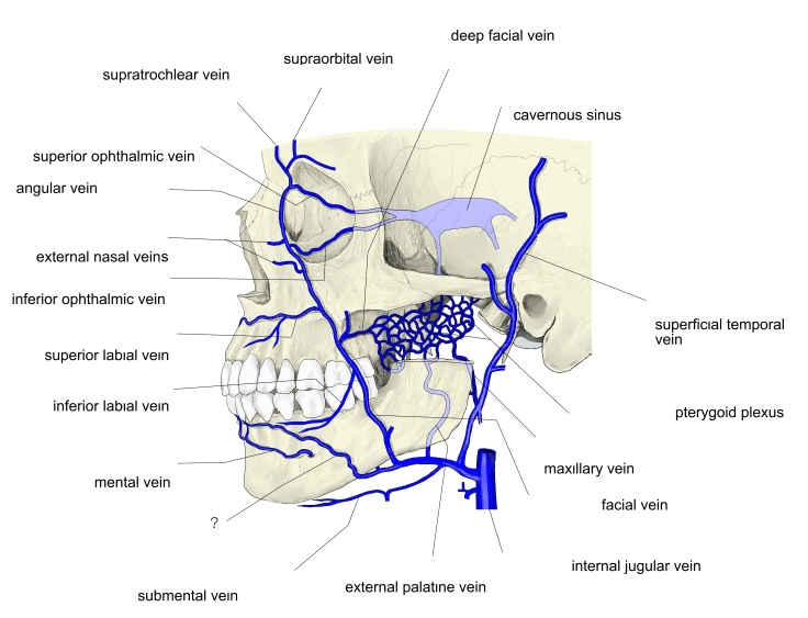
Veins of the face
Main venous plexuses
| Plexus | Position | Main outflow |
|---|---|---|
| Pterygoid plexus | Infratemporal fossa, around the pterygoid muscles | Maxillary vein → retromandibular vein |
| Pharyngeal plexus | Outer surface of the pharynx | Internal jugular vein |
| Basilar plexus | On the clivus, inside the skull | Petrosal sinuses, internal vertebral plexus |
| Suboccipital plexus | Suboccipital region | Vertebral and deep cervical veins |
| Internal and external vertebral venous plexuses | Inside and outside the vertebral canal | Intervertebral veins → segmental veins |
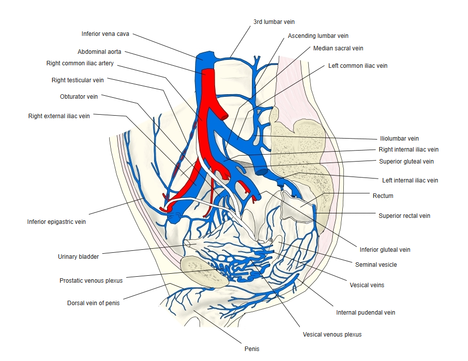
| Vesical plexus | Base of the bladder | Internal iliac veins |
| Rectal plexus | Wall of the rectum and anal canal | Superior, middle and inferior rectal veins |
| Uterine plexus / vaginal plexus | Beside the uterus and vagina | Internal iliac veins |
| Prostatic plexus | Around the prostate | Internal iliac veins |
| Pampiniform plexus | Spermatic cord | Testicular vein |
Veins of the male pelvis
Clinical notes
- Valveless plexuses such as the vertebral venous plexus can carry infection or tumour cells between regions (for example from the pelvis to the vertebrae).
- Dilated plexuses form varicoceles (pampiniform plexus) and haemorrhoids (rectal plexus).
References and sources
- Further reading: Venous plexus, Wikipedia.