Cuneiform bones (Latin: ossa cuneiformia)
Also called Cuneiforms
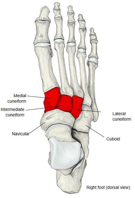
One group of the tarsal bones of the foot. There are three of them, from medial to lateral the medial cuneiform, the intermediate cuneiform and the lateral cuneiform, lying in a row between the navicular and the first three metatarsal bones. The name is usually used with one of the words medial, intermediate or lateral rather than simply as "cuneiform bone".
bones
- Distal tarsal bones
- Cuneiform bones : from the big-toe side to the little-toe side: medial cuneiform, intermediate cuneiform, lateral cuneiform.
- Cuboid : on the little-toe side.
- Proximal tarsal bones : navicular, talus, calcaneus.
· The intermediate and lateral cuneiforms are narrower on the plantar side than on the dorsal side; this wedge shape helps to form the transverse arch of the foot. The cuneiforms articulate with each other at the intercuneiform joints, with the navicular behind at the cuneonavicular joint and with the metatarsals in front.
Reference websites
[Wikipedia]
· Cuneiform bones, Wikipedia.
[Websites with illustrations]
· Bones of the foot, lateral view (Gray's Anatomy) (Wikimedia Commons)

· Cuneiform bone (drawing) (ClipArt ETC (University of South Florida))

· Cuneiform bones: anatomy and clinical notes (Kenhub)
Related terms
- Medial cuneiform
- Intermediate cuneiform
- Lateral cuneiform
- Tarsal bones
- Distal tarsal bones
- Intercuneiform joints
- Cuneonavicular joint
References and sources
- Further reading: Cuneiform bones, Wikipedia.
- Federative International Programme for Anatomical Terminology (FIPAT). Terminologia Anatomica, 2nd ed. (TA2), 2019.