Latin: os cuneiforme laterale · also called the third cuneiform
Overview
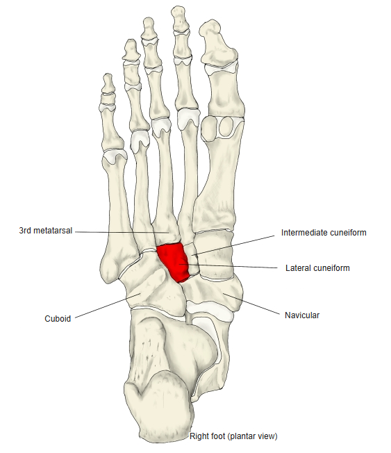
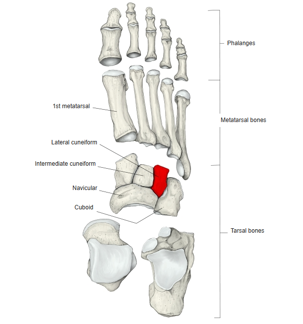
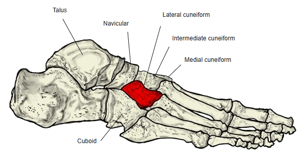
- One of the seven tarsal bones; it is the most lateral of the three cuneiforms and the second largest, after the medial cuneiform.
- It is shaped like a wedge: viewed from the front it is triangular, with its apex directed downwards (towards the sole).
- It has articular surfaces on its anterior, posterior, medial and lateral sides.
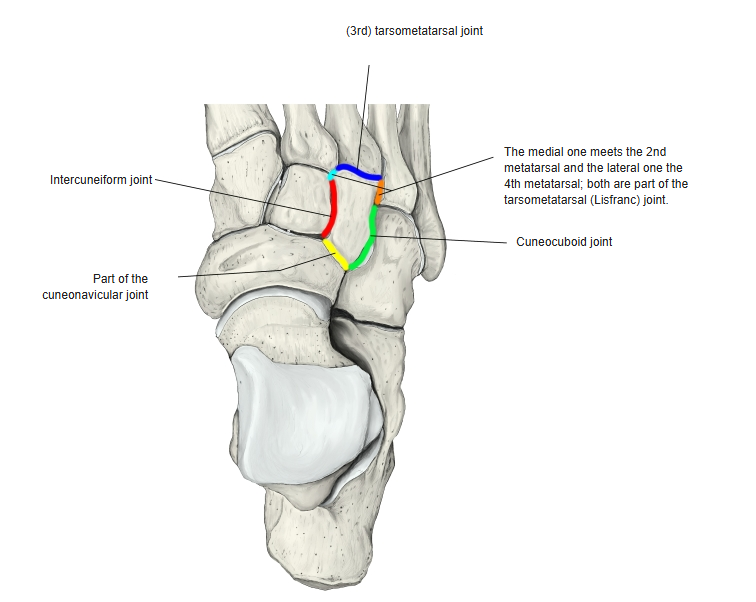
- It articulates with six bones: the 2nd, 3rd and 4th metatarsals, the intermediate cuneiform, the cuboid and the navicular.
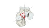
Shape of the lateral cuneiform
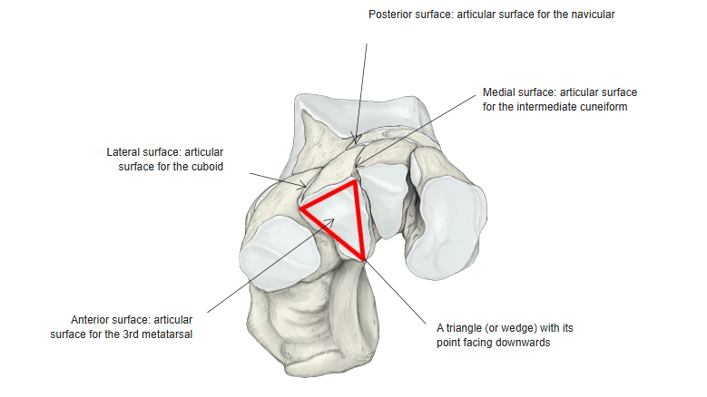
- Bones of the lower limb (one side)
- Pelvic girdle
- Hip bone ← ilium, ischium and pubis fused
- Free lower limb
- Thigh and knee
- Femur
- Patella
- Leg
- Tibia ← big-toe side
- Fibula ← little-toe side
- Foot
- Tarsal bones (7)
- Metatarsal bones (5)
- Phalanges (14)
- Thigh and knee
- Pelvic girdle

Illustrations
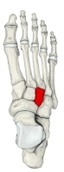
Right foot (dorsal) 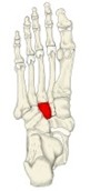
Right foot (plantar) 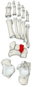
Bones of the foot (separated) 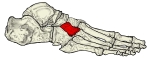
Right foot (lateral) 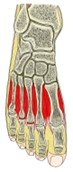
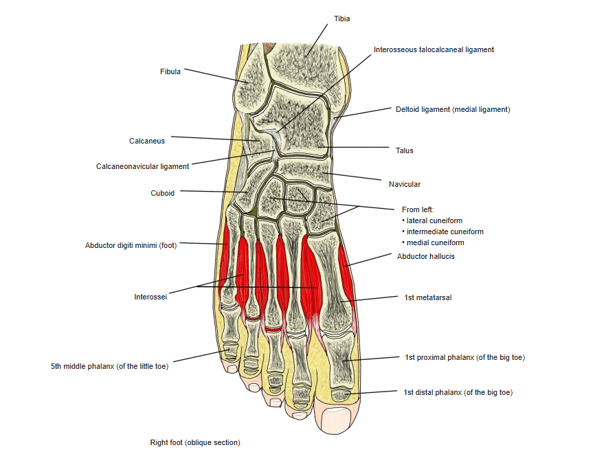
Right foot (oblique section) 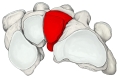
Distal tarsals and metatarsals (posterior) 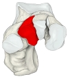
Tarsal bones (anterior) 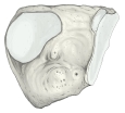
Right lateral cuneiform (lateral surface) 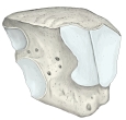
Right lateral cuneiform (medial surface)
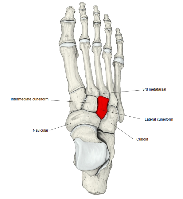
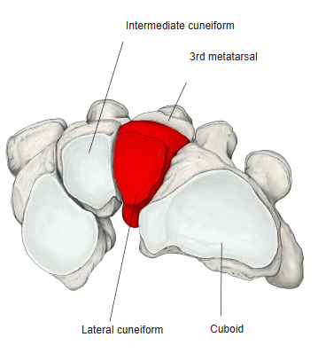
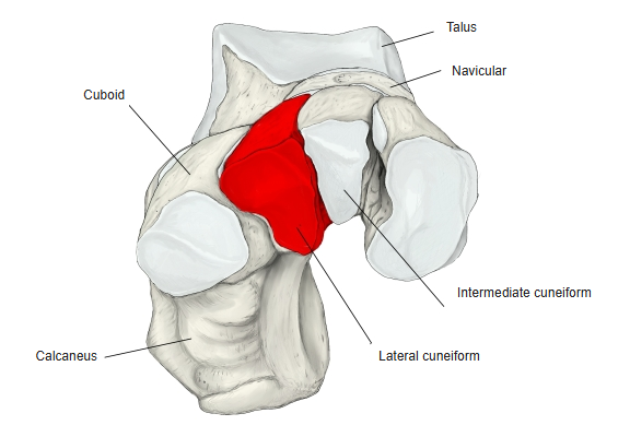
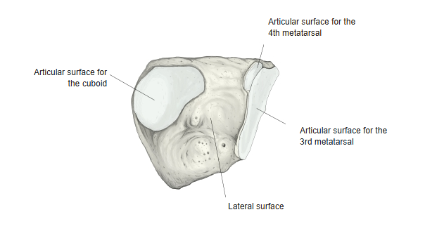
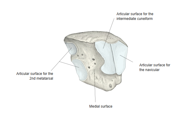
Articulations
| # | Bone | Joint |
|---|---|---|
| 1 | 2nd metatarsal | Tarsometatarsal joint |
| 2 | 3rd metatarsal | |
| 3 | 4th metatarsal | |
| 4 | Intermediate cuneiform | Intercuneiform joint |
| 5 | Cuboid | Cuneocuboid joint |
| 6 | Navicular | Cuneonavicular joint |
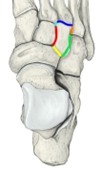
Joints of the lateral cuneiform
Ligaments
The following ligaments attach to the lateral cuneiform. The list is intended to be comprehensive but may omit minor ligaments.
| # | Ligament | Connects |
|---|---|---|
| 1 | Dorsal tarsometatarsal ligaments | Lateral cuneiform – metatarsals |
| 2 | Plantar tarsometatarsal ligaments | Lateral cuneiform – metatarsals |
| 3 | Interosseous cuneometatarsal ligament | Lateral cuneiform – 4th metatarsal |
| 4 | Dorsal intercuneiform ligament | Lateral cuneiform – intermediate cuneiform |
| 5 | Plantar intercuneiform ligament | Lateral cuneiform – intermediate cuneiform |
| 6 | Interosseous intercuneiform ligament | Lateral cuneiform – intermediate cuneiform |
| 7 | Dorsal cuneocuboid ligament | Lateral cuneiform – cuboid |
| 8 | Plantar cuneocuboid ligament | Lateral cuneiform – cuboid |
| 9 | Dorsal cuneonavicular ligament | Lateral cuneiform – navicular |
| 10 | Plantar cuneonavicular ligament | Lateral cuneiform – navicular |
Muscle attachments
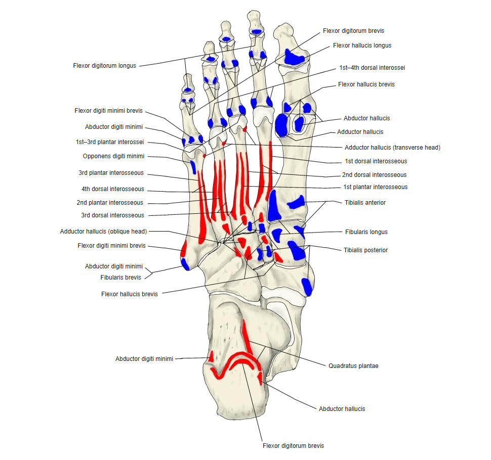
- Origin of: Flexor hallucis brevis, Adductor hallucis (oblique head)
- Insertion of: Tibialis posterior
All of these attachments are on the plantar surface.
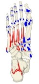
Muscle attachments on the sole (red: origin, blue: insertion)
References and sources
- Further reading: Lateral cuneiform bone, Wikipedia.