Dental pulp (Latin: pulpa dentis)
Also called the tooth pulp
- Overview
- Divisions
- Structure
- Reference websites
- (Wikipedia, websites with illustrations)
- Where it is found
- Related terms
Overview
· It develops from the dental papilla of the tooth germ, a mesenchyme of neural crest origin (ectomesenchyme).
· Its outline roughly follows the outer shape of the dentin, and so of the tooth.
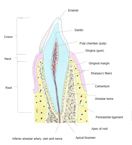
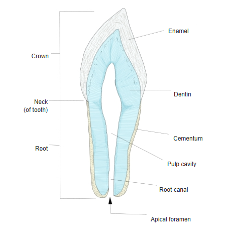
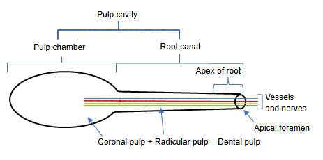
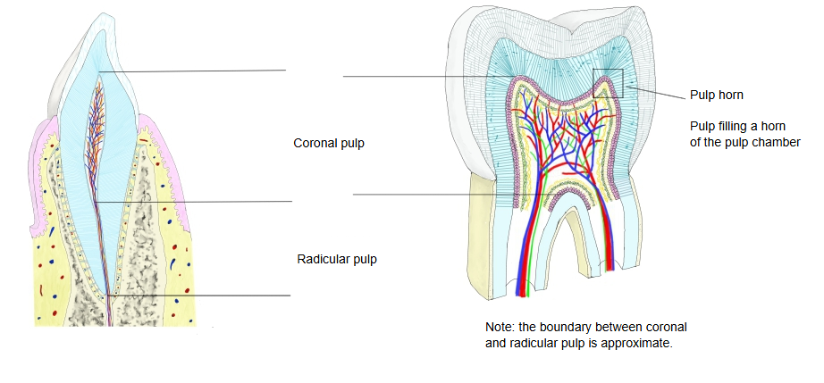
Vessels and nerves enter through the apical foramen and run up the root canal; they come from the alveolar arteries and from the superior and inferior dental plexuses. Pulp and dentin form one functional unit, the pulp–dentin complex: the pulp forms and nourishes the dentin, senses stimuli (felt mainly as pain) and defends the tooth. The pulp becomes smaller with age, because dentin keeps being laid down on the walls of the pulp cavity. (Sources differ on where the coronal pulp ends and the radicular pulp begins; the boundary is usually placed at the level of the neck of the tooth.)
a tooth
- Hard tissues
- Dental pulp
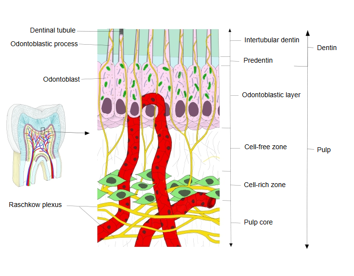
- Vessels and nerves : arteries, veins, lymph vessels and nerves (which form the plexus of Raschkow) are plentiful.
- Cells : odontoblasts in the outermost layer; also fibroblasts, undifferentiated mesenchymal cells and others.
- Connective tissue : mainly collagen fibres (types I and III); elastic fibres only in the walls of the larger vessels.
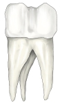 Parts of a tooth
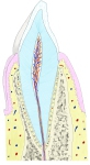 Tooth in its socket (section)
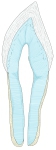 Hard tissues of a tooth (section)
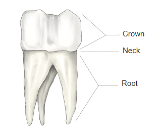
Divisions
· The extensions of the coronal pulp that fill the pulp horns of the chamber, under the cusps, are themselves called pulp horns.
· Both blood vessels and nerves are especially well developed in the pulp horns.
Structure
of the pulp
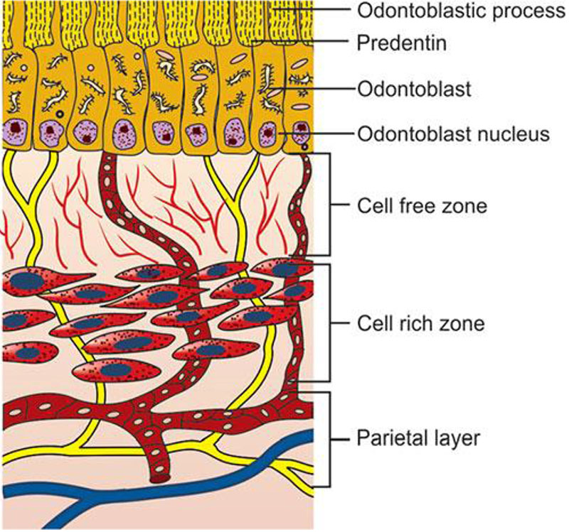
- Odontoblastic layer : the outermost layer: a single row of odontoblasts, whose long processes run into the dentin (the dentinal fibres); rich in capillaries.
- Cell-free zone : few cells (zone of Weil); often hard to see in the radicular pulp. Rich in capillaries and nerve fibres: the plexus of Raschkow lies here.
- Cell-rich zone : many fibroblasts and undifferentiated mesenchymal cells.
- Pulp core : everything inside the three layers above; it forms most of the pulp and carries the larger vessels and nerves.
Blood vessels are plentiful from the odontoblastic layer to the cell-free zone. Under the odontoblasts the nerve fibres form the subodontoblastic plexus (of Raschkow); some of its branches enter the dentinal tubules and reach the odontoblast processes.
The pulp contains many kinds of cells: odontoblasts, which form dentin from the pulp side throughout life; fibroblasts (pulp cells), which make and break down the collagen fibres; and undifferentiated mesenchymal cells (dental pulp stem cells) near the vessels, which can become odontoblasts or fibroblasts. Immune cells include macrophages, lymphocytes and dendritic cells; neutrophils appear in inflammation of the pulp (pulpitis).
Reference websites
[Wikipedia]
· Pulp (tooth), Wikipedia.
· Dentin, Wikipedia.
[Websites with illustrations]
· Histology of the pulp (stock illustration) (iStock)

· Pulp zones (The Future Dentistry)
Where it is found
Related terms
- Pulp cavity
- Root canal
- Periodontal ligament
- Teeth
- Superior dental plexus
- Inferior dental plexus
- Inferior alveolar nerve
References and sources
- Further reading: Pulp (tooth), Wikipedia.
- Further reading: Dentin, Wikipedia.
- Federative International Programme for Anatomical Terminology (FIPAT). Terminologia Anatomica, 2nd ed. (TA2), 2019.