Periodontal ligament
Also called the periodontal membrane
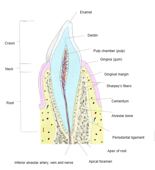
The periodontal ligament is the thin layer of dense fibrous connective tissue between the cementum of a tooth root and the bone of its socket, which holds the tooth in place. It is one of the four periodontal tissues (periodontium), with the gingiva, the cementum and the alveolar bone of the alveolar process. It is only about 0.15–0.4 mm wide, thinnest around the middle third of the root, and becomes thinner with age.
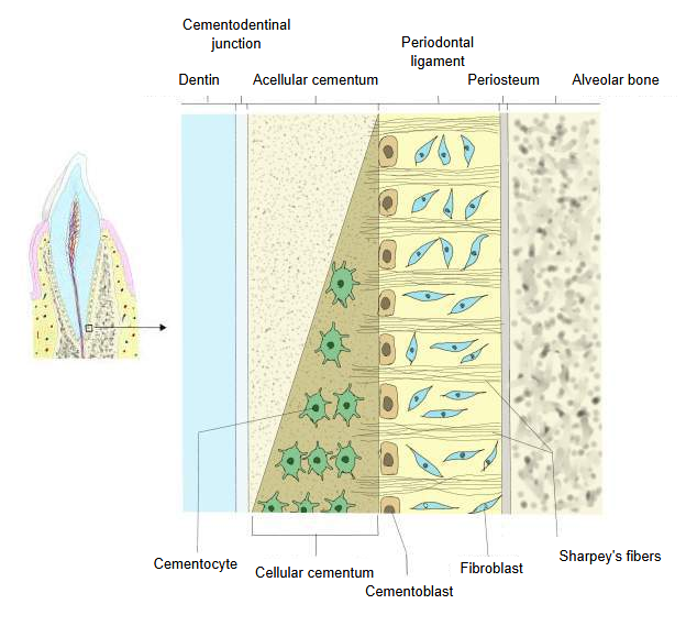
Its collagen fibre bundles are anchored in the cementum on one side and in the alveolar bone on the other (the embedded ends are Sharpey's fibres), so the tooth is suspended in its socket rather than fused to the bone: a fibrous joint called a gomphosis (dentoalveolar joint). The fibre groups are the alveolar crest, horizontal, oblique (the most numerous, taking most of the chewing load), apical and, in multirooted teeth, interradicular fibres; the transseptal fibres, which run over the bone crest from tooth to tooth, are often counted as gingival fibres.
Its main cells are fibroblasts, with cementoblasts on the root side, osteoblasts and osteoclasts on the bone side, undifferentiated stem cells for repair and the epithelial cell rests of Malassez around the root. Loose connective tissue between the fibre bundles carries the vessels and nerves (from the apical vessels, vessels perforating the socket wall and the gingiva); its mechanoreceptors sense the force and direction of biting, and its cells remodel the bone, as in orthodontic tooth movement.
Sources differ on the name: periodontal ligament and periodontal membrane usually mean the same tissue, but some older texts use 'periodontal ligament' only for the strong ring of fibres around the neck of the tooth at the edge of the socket. See also the Teeth page, the root canal and the dental pulp.
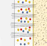 Periodontal ligament (diagram)
 Cementum and periodontal ligament (microscopic)
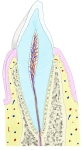 Tooth in its socket (section)
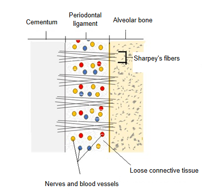
Where it is found
Related terms
References and sources
- Further reading: Periodontal ligament, Wikipedia.
- Further reading: Periodontium, Wikipedia.
- Federative International Programme for Anatomical Terminology (FIPAT). Terminologia Anatomica, 2nd ed. (TA2), 2019.