Falx cerebri (Latin: falx cerebri)
Also called the cerebral falx
What is the falx cerebri?
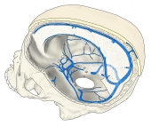 Falx cerebri in the opened skull
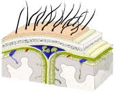 Scalp, skull and meninges (coronal)
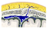 Superior sagittal sinus (coronal)
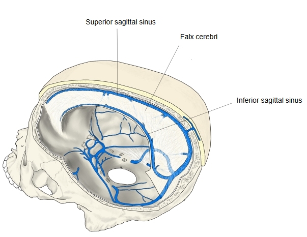
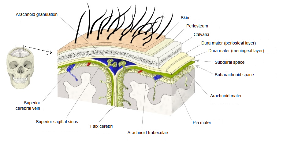
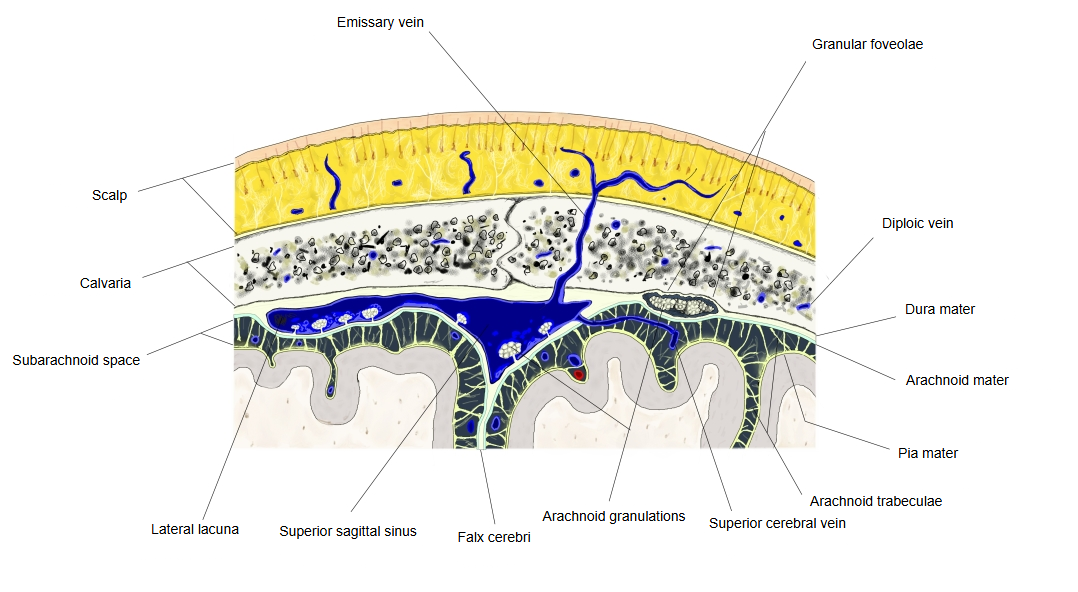
The falx cerebri is a sickle-shaped fold of dura mater that runs from front to back along the midline of the skull, with its cutting edge facing down. It is formed by the inner (meningeal) layer of the dura mater, folded double. It reaches deep into the longitudinal fissure and separates the two cerebral hemispheres, but does not reach the corpus callosum. In front it arises from the crista galli of the ethmoid bone; it runs back along the groove for the superior sagittal sinus to the internal occipital protuberance of the occipital bone, and continues into the upper surface of the tentorium cerebelli. Its attached upper border splits into two layers that enclose the superior sagittal sinus; its free, sickle-shaped lower border also splits into two layers and contains the inferior sagittal sinus.
From the crista galli, the upper border runs back along the frontal crest and the groove for the superior sagittal sinus. The free lower border runs in the longitudinal fissure a little above the corpus callosum and contains the narrow inferior sagittal sinus. Behind and below, the falx is fused with the upper surface of the tentorium cerebelli; the line of fusion contains the straight sinus. These sinuses belong to the dural venous sinuses.
The name comes from its sickle-like form (Latin falx, sickle); it is also called the cerebral falx. It limits side-to-side movement of the brain; a mass on one side can push the cingulate gyrus under its free edge (subfalcine herniation).
Reference websites
[Wikipedia]
· Falx cerebri, Wikipedia.
· Dura mater, Wikipedia.
[Websites with illustrations]
· Falx cerebri in a dissected head, medial view (specimen photograph) (Tumblr)

Video
Where it is found
Related terms
- Dura mater
- Crista galli
- Superior sagittal sinus
- Inferior sagittal sinus
- Straight sinus
- Granular foveolae
References and sources
- Further reading: Falx cerebri, Wikipedia.
- Further reading: Dura mater, Wikipedia.
- Federative International Programme for Anatomical Terminology (FIPAT). Terminologia Anatomica, 2nd ed. (TA2), 2019.