Latin: Sinus durae matris
Overview
- Venous channels between the two layers of the dura mater (periosteal and meningeal) or in its folds.
- They have no valves and no muscle in their walls; their walls are dura lined by endothelium.
- They receive blood from the brain, the meninges and the skull bones (diploic veins), and drain mainly into the internal jugular vein.
- They connect with veins outside the skull through the emissary veins.
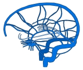
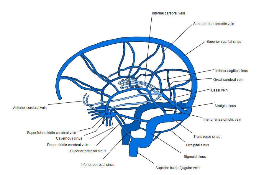
Dural venous sinuses and cerebral veins 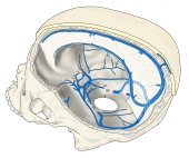
Dural venous sinuses in the skull
Groups
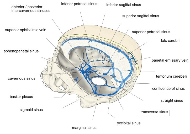
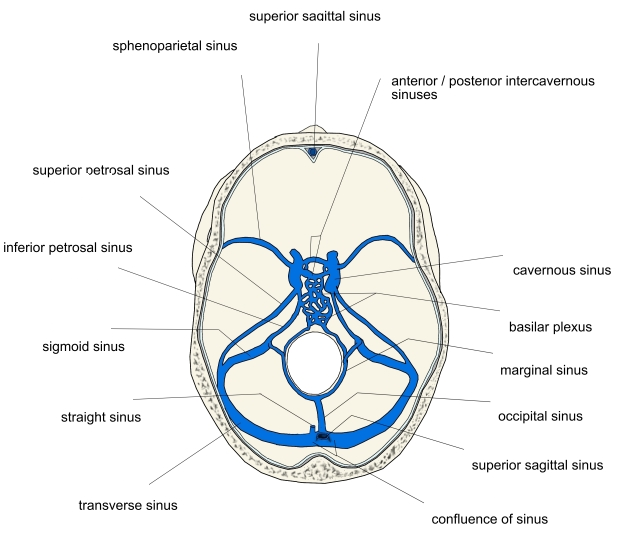
- Posterosuperior group (mostly in the midline and its continuations): superior sagittal, inferior sagittal, straight, occipital sinuses, the confluence of sinuses, transverse and sigmoid sinuses.
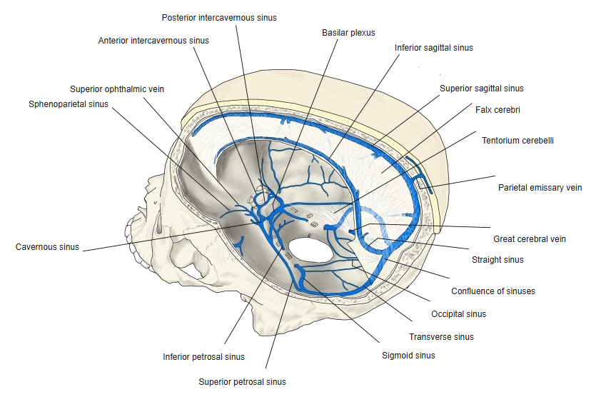
- Antero-inferior group (paired, around the cavernous sinus on the cranial base): cavernous, anterior and posterior intercavernous, sphenoparietal, superior and inferior petrosal sinuses, and the basilar plexus.
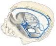
Dural venous sinuses (side view) 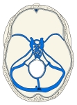
Dural venous sinuses of the cranial base
Drainage
- Blood flows backward along the roof and midline to the confluence of sinuses, then laterally through the transverse and sigmoid sinuses to the jugular foramen.
- The cavernous sinuses drain backward through the petrosal sinuses to the transverse–sigmoid junction and the internal jugular vein.
Clinical notes
- Dural venous sinus thrombosis can cause headache, raised intracranial pressure and venous infarcts of the brain.
- Infection can spread from the face and scalp into the sinuses through valveless emissary and ophthalmic veins.
References and sources
- Further reading: Dural venous sinuses, Wikipedia.