Osseous labyrinth
Also called the bony labyrinth or the otic capsule
The osseous labyrinth (bony labyrinth) is the outer part of the labyrinth: the part where calcium salts were laid down in the cartilage around it, so that it became bone forming the walls of tubes.
Labyrinth: another name for the internal ear (part of the organ of hearing), named for its complex shapeWall thickness: about 2–3 mm
· The walls of its tubes are only about 2–3 mm thick. Its long axis is about 2 cm and runs almost parallel to the axis of the petrous part of the temporal bone.
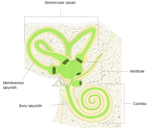
· The membranous labyrinth is much narrower than the bony labyrinth. Part of its wall is attached to the bony wall, but most of it lies apart from the bone, leaving a wide space filled with perilymph.
Inside, it holds the membranous labyrinth, and the space between the two is filled with perilymph.
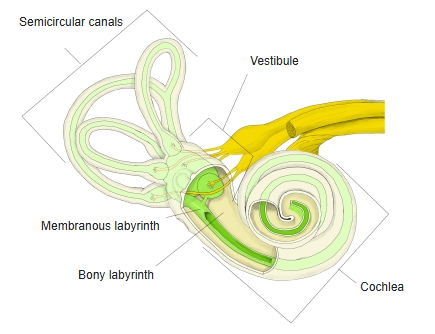
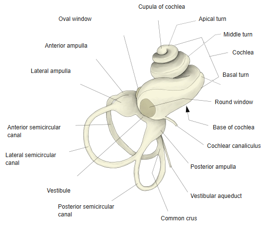
The two labyrinths have almost the same shape; their main parts are three: the cochlea, the vestibule and the semicircular canals.
The vestibule differs considerably between the twoCochlea: organ of hearingSemicircular canals (and vestibule): organ of balance
- Osseous (bony) labyrinth : the walls of its tubes are about 2–3 mm thick, formed where calcium salts were laid down in cartilage and it became bone. It holds the membranous labyrinth; the space between the two is filled with perilymph.
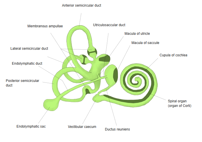
- Membranous labyrinth : fine membranous tubes of almost the same shape as the bony labyrinth, filled with endolymph.Main parts of both labyrinths: cochlea, vestibule, semicircular canals.
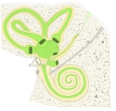 Bony and membranous labyrinth
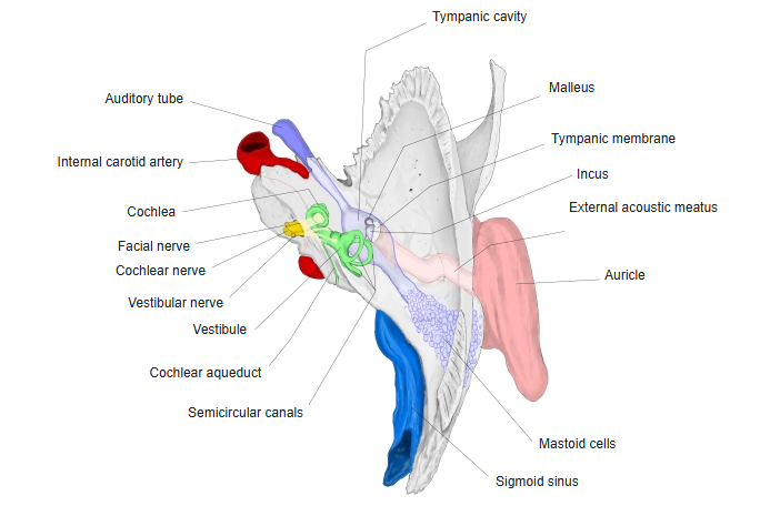
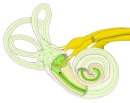 Labyrinth and nerves
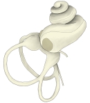 Bony labyrinth
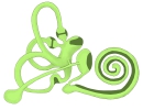 Membranous labyrinth
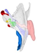 Temporal bone (superior view)
Reference websites
[Wikipedia]
· Bony labyrinth, Wikipedia.
[Websites with illustrations]
· Right osseous labyrinth, lateral view (Gray's Anatomy, fig. 920) (Wikimedia Commons)

· Interior of the right osseous labyrinth (Gray's Anatomy, fig. 921) (Wikimedia Commons)

· The inner ear: bony and membranous labyrinth (TeachMeAnatomy)