Latin: auris interna
Overview
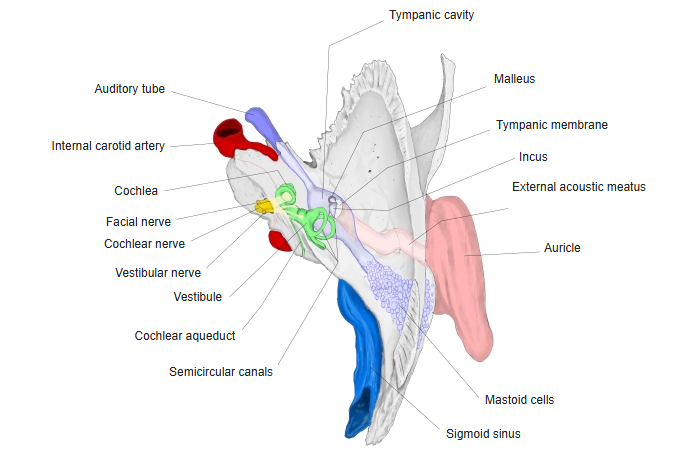
- The innermost part of the ear, in the petrous part of the temporal bone; it contains the receptors for hearing and balance.
- Also called the labyrinth because of its complicated shape.
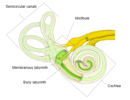
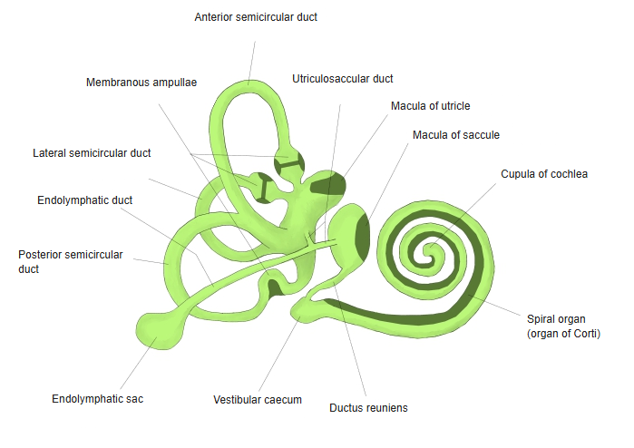
- A system of bony cavities, the bony labyrinth, contains a system of membranous ducts and sacs, the membranous labyrinth.
- The space between them holds perilymph; the membranous labyrinth is filled with endolymph.
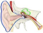
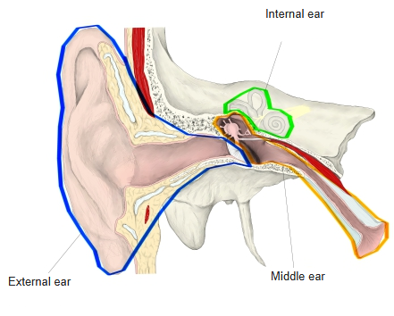
External, middle and internal ear 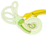
Bony and membranous labyrinth
Parts
| Bony labyrinth | Membranous labyrinth inside | Function |
|---|---|---|
| Cochlea | Cochlear duct with the spiral organ | Hearing |
| Vestibule | Utricle and saccule with their maculae | Balance: head position and linear acceleration |
| Semicircular canals | Semicircular ducts with the ampullary crests | Balance: rotation of the head |
- The oval and round windows in the medial wall of the tympanic cavity open into the vestibule and the cochlea.
- Nerves: the vestibulocochlear nerve enters through the internal acoustic meatus.
- Blood supply: the labyrinthine artery, usually from the anterior inferior cerebellar artery.
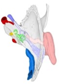
Right ear in the temporal bone (superior view) 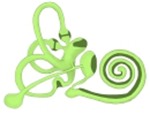
Membranous labyrinth
References and sources
- Further reading: Internal ear, Wikipedia.