Reticular cell (Latin: Cellula reticularis)
· Reticular tissue forms the supporting network of soft organs such as the liver and the bone marrow, and of the tissues and organs of the lymphatic system, such as the lymphoid nodules, lymph nodes and the spleen. The framework of the thymus is made by epithelial reticular cells (see below).
In more detail
Reticular cells produce the reticular fibres and wrap them in their cytoplasm, separating the fibres from the other components of the tissue and from the free cells around them. They are found in many tissues, including the spleen, lymph nodes and lymphoid nodules. Reticular fibres are also made by fibroblasts; reticular cells are in fact a specialised kind of fibroblast and synthesise the type III collagen of the fibres. In lymphoid tissue, reticular cells guide B lymphocytes and T lymphocytes to their own zones.
The resulting mesh of cells and fibres (the stroma) holds lymphocytes, macrophages and blood-forming cells in place. In lymphoid organs these cells are called fibroblastic reticular cells. The epithelial reticular cells of the thymus come from epithelium rather than mesenchyme; sources differ on whether to call them reticular cells.
Reference websites
[Wikipedia]
· Reticular cell, Wikipedia.
· Reticular connective tissue, Wikipedia.
[Websites with illustrations]
· Reticular cells and their processes in a lymph node (micrograph) (Anatomy Atlases)
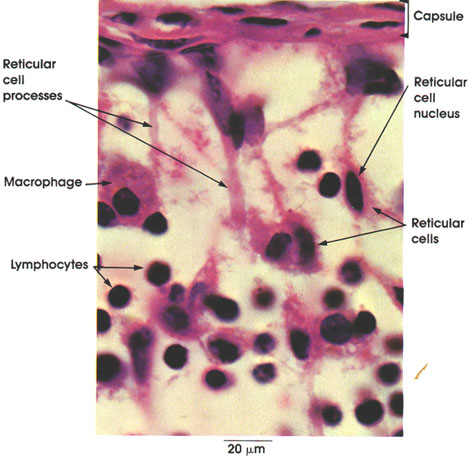
· Reticular cells among lymphocytes (micrograph) (Loyola University Chicago (LUMEN))

· Fibroblastic reticular cell network of the lymph node (figure) (Nature Reviews Immunology)

Related terms
- Fibroblast
- Reticular fibres
- Connective tissue proper
- Stromal cell
- Lymph node
- Spleen
- Red bone marrow
- Lymphoid organ
References and sources
- Further reading: Reticular cell, Wikipedia.
- Further reading: Reticular connective tissue, Wikipedia.
- Federative International Programme for Anatomical Terminology (FIPAT). Terminologia Anatomica, 2nd ed. (TA2), 2019.