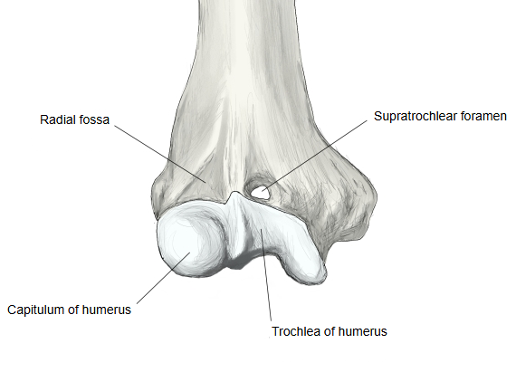
Supratrochlear foramen (Latin: foramen supratrochleare)
Also called the septal aperture or the intercondylar foramen
It forms when the thin plate of bone that normally separates the two fossae is absent; in life the opening is closed by a thin membrane. It is also called the septal aperture. It is an anatomical variation, not a regular feature: it has no Terminologia Anatomica name, and its frequency differs widely between populations. Sources differ, but it is reported more often in women and on the left side. In some mammals, for example dogs, it is a normal feature. On X-rays it can look like a cyst or a lytic lesion, and it may make the humerus narrower at this level.
Reference websites
[Wikipedia]
· Supratrochlear foramen, Wikipedia.
Sites with illustrations
· Variation in the septal aperture of the distal humerus (photographs, slide)

Where it is found
Related terms
- Coronoid fossa
- Olecranon fossa
- Radial fossa
- Trochlea of humerus
- Supracondylar process
- Lower end of humerus
References and sources
- Further reading: Supratrochlear foramen, Wikipedia.
- Federative International Programme for Anatomical Terminology (FIPAT). Terminologia Anatomica, 2nd ed. (TA2), 2019.