Latin: humerus · the bone of the arm
Overview
- The only bone of the arm — a long bone running from the shoulder joint to the elbow joint.
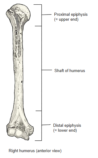
- It has an upper end (proximal epiphysis), a shaft and a lower end (distal epiphysis).
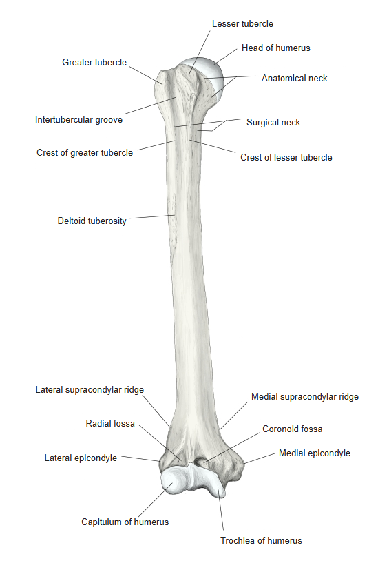
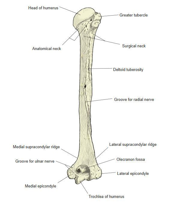
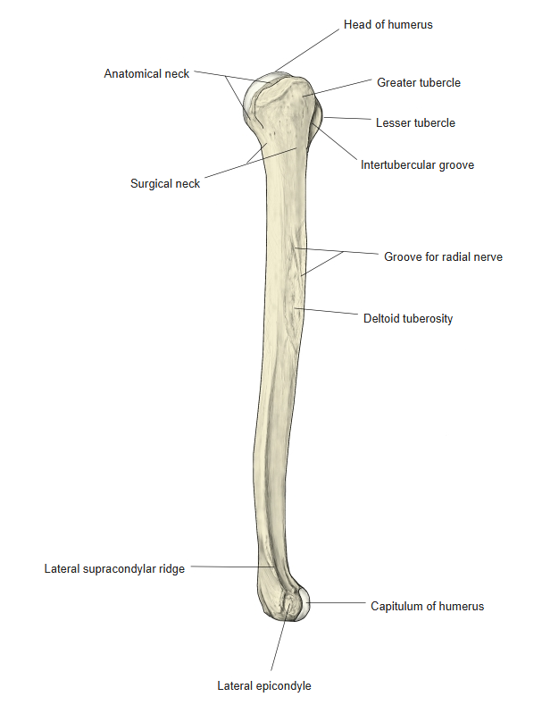
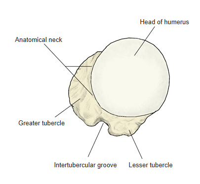
- Upper end: the rounded head of the humerus, the anatomical neck, the greater and lesser tubercles with the intertubercular groove between them, and the surgical neck below.
- Shaft: the deltoid tuberosity on the lateral side and the groove for the radial nerve on the posterior surface.
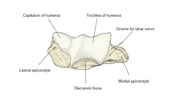
- Lower end: the capitulum (for the radius) and the trochlea (for the ulna), the medial and lateral epicondyles, and the coronoid, radial and olecranon fossae.
Right humerus 
Parts of the humerus
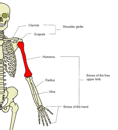
- Bones of the upper limb (one side)
- Shoulder girdle
- Clavicle
- Scapula
- Free upper limb
- Arm
- Humerus
- Forearm
- Radius ← thumb side
- Ulna ← little-finger side
- Hand
- Carpal bones (8)
- Metacarpal bones (5)
- Phalanges (14)
- Arm
- Shoulder girdle
Illustrations
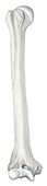
Right humerus (anterior) 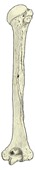
Right humerus (posterior) 
Right humerus (lateral) 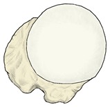
Upper end, seen from above 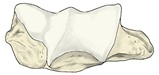
Lower end, seen from below
Articulations
| # | Bone | Joint |
|---|---|---|
| 1 | Scapula | Glenohumeral (shoulder) joint |
| 2 | Ulna | Humeroulnar joint |
| 3 | Radius | Humeroradial joint |
Joints of the humerus
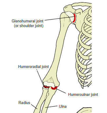
Glenohumeral joint: head of the humerus ↔ glenoid cavity of the scapula. Humeroulnar joint: trochlea of the humerus ↔ trochlear notch of the ulna. Humeroradial joint: capitulum of the humerus ↔ articular fovea on the head of the radius.
Ligaments
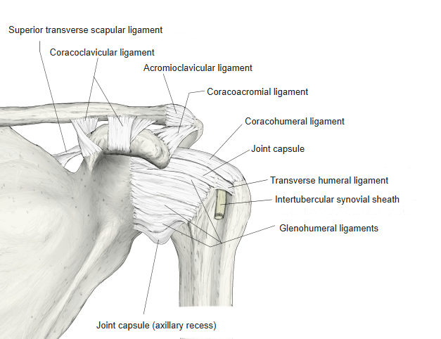
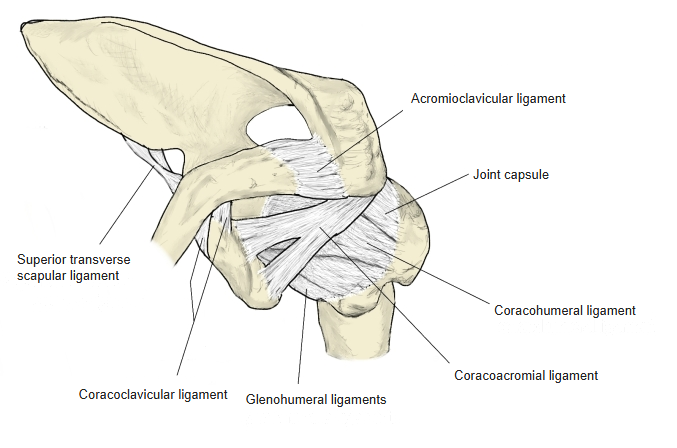
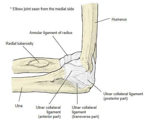
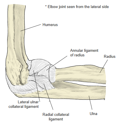
The following ligaments attach to the humerus. The list is intended to be comprehensive but may omit minor ligaments.
| # | Ligament | Connects |
|---|---|---|
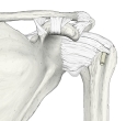
| 1 | Coracohumeral ligament | Greater and lesser tubercles – coracoid process of scapula |
| 2 | Glenohumeral ligaments | Glenoid labrum – anatomical neck |
| 3 | Transverse humeral ligament | Between the greater and lesser tubercles (over the intertubercular groove) |
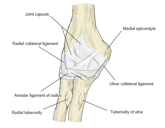
| 4 | Ulnar collateral ligament of the elbow (anterior, posterior and transverse parts) | Medial epicondyle – coronoid process and olecranon of the ulna (the transverse part runs between the coronoid process and the olecranon) |
| 5 | Radial collateral ligament of the elbow | Lateral epicondyle – annular ligament of the radius (some fibres reach the ulna) |
| 6 | Struthers' ligament (when present) | Medial epicondyle – supracondylar process |
Right shoulder joint (anterior) 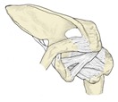
Right shoulder joint (from above) 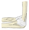
Right elbow joint (medial) 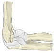
Right elbow joint (lateral) 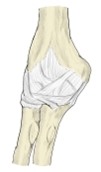
Right elbow joint (posterior)
Muscle attachments
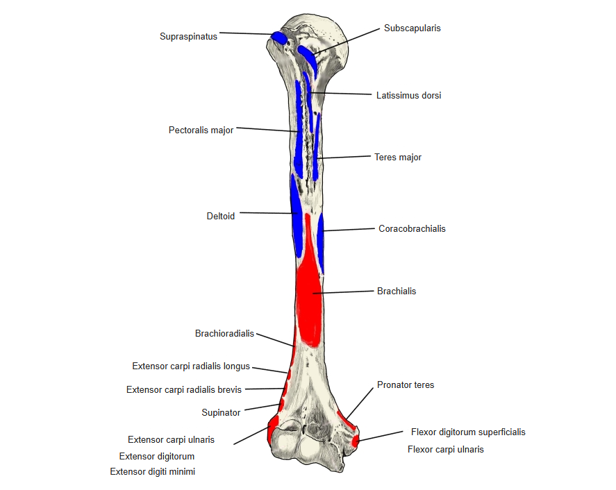
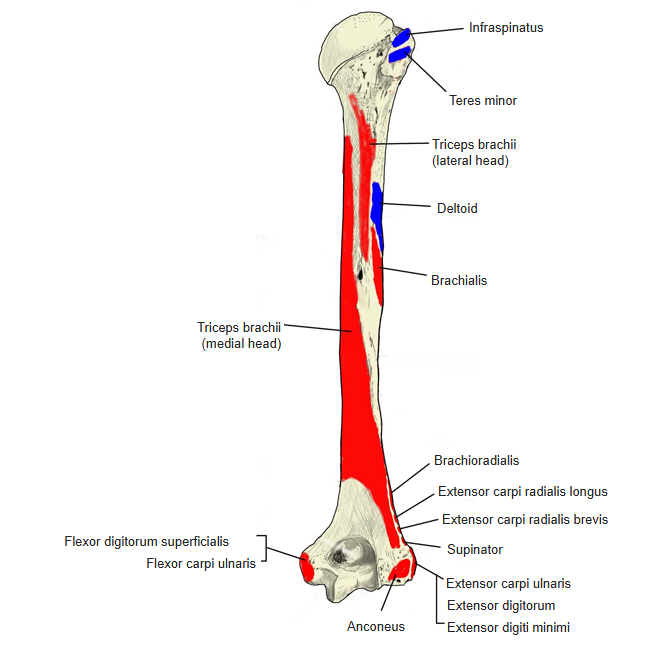
- Origin of: Brachialis, Triceps brachii, Anconeus, Supinator, Brachioradialis, Pronator teres, Extensor carpi radialis longus, Extensor carpi radialis brevis, Extensor digitorum, Extensor digiti minimi, Extensor carpi ulnaris, Flexor carpi radialis, Palmaris longus, Flexor carpi ulnaris, Flexor digitorum superficialis
- Insertion of: Supraspinatus, Infraspinatus, Teres minor, Subscapularis, Teres major, Coracobrachialis, Deltoid, Pectoralis major, Latissimus dorsi
Origins: brachialis — anterior surface, lower half; triceps brachii (lateral and medial heads) — posterior surface; anconeus, supinator, extensor carpi radialis brevis, extensor digitorum, extensor digiti minimi and extensor carpi ulnaris — lateral epicondyle; brachioradialis — upper two-thirds of the lateral supracondylar ridge; extensor carpi radialis longus — lower third of the lateral supracondylar ridge; pronator teres (humeral head), flexor carpi radialis, palmaris longus, flexor carpi ulnaris (humeral head) and flexor digitorum superficialis (humeroulnar head) — medial epicondyle (common flexor origin). Insertions: supraspinatus, infraspinatus and teres minor — greater tubercle; subscapularis — lesser tubercle (and its crest); teres major — crest of lesser tubercle; latissimus dorsi — floor of the intertubercular groove; pectoralis major — crest of greater tubercle; deltoid — deltoid tuberosity; coracobrachialis — medial side of the shaft, middle.
Muscle attachments (anterior) 
Muscle attachments (posterior)
Borders and surfaces
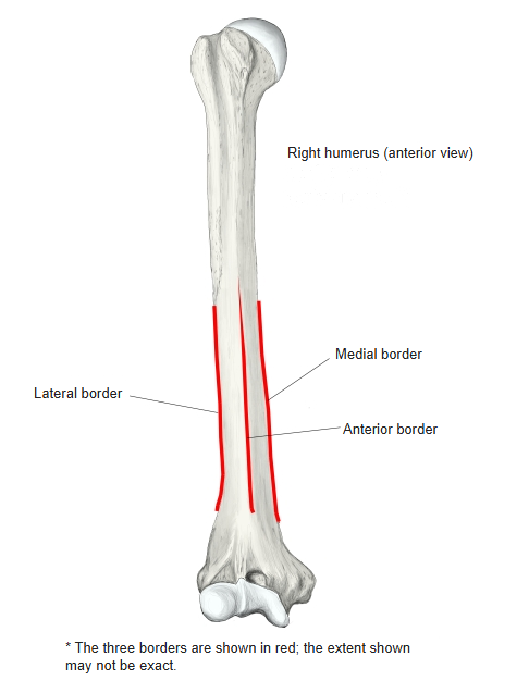
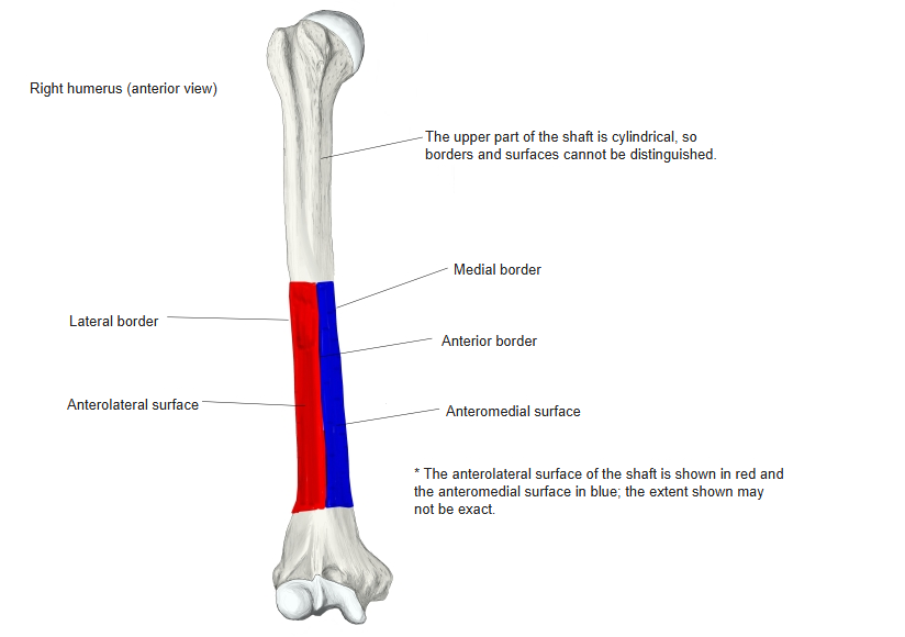
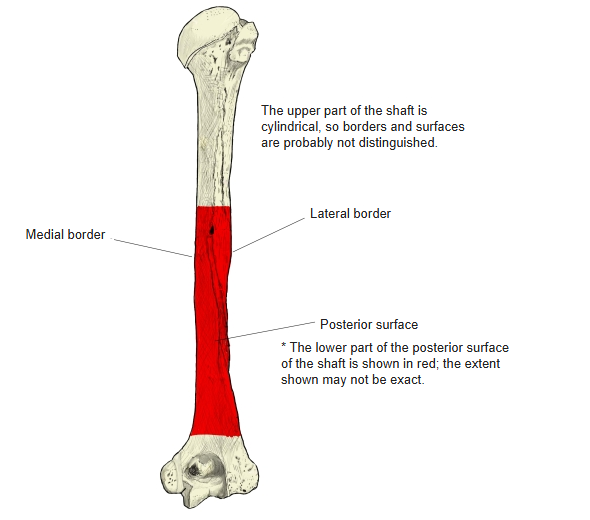
- Seen in the lower half of the shaft; the upper part is cylindrical, so borders and surfaces are not distinguished there.
- Borders: anterior, medial and lateral. The anterior border is rounded; the medial and lateral borders become sharp near the lower end.
- Surfaces: anteromedial, anterolateral and posterior. Many muscles arise from or attach to these surfaces.
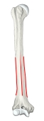
Borders 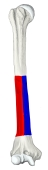
Surfaces (anterior) 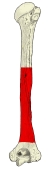
Posterior surface
Development
- The ossification centre of the shaft appears at about the 8th fetal week.
- Further centres appear in the head (1–2 years), capitulum (about 2 years), greater tubercle (2–3 years), lesser tubercle (3–5 years), medial epicondyle (5–8 years), trochlea (11–12 years) and lateral epicondyle (12–14 years); they fuse to form the two ends.
- The ends fuse with the shaft at about 16–25 years.
Anomalies
- Supracondylar process: a hook-like projection sometimes found above the medial epicondyle.
- A ligament (Struthers' ligament) may run from its tip to the medial epicondyle, forming a foramen for the median nerve; part of the pronator teres may arise from it. The ligament may ossify into a bony bridge (normal in some animals).
- Supratrochlear foramen: the thin bony plate between the olecranon fossa and the coronoid fossa may be perforated.
References and sources
- Further reading: Humerus, Wikipedia.