Tarsal glands (Latin: glandulae tarsales)
Also called Meibomian glands
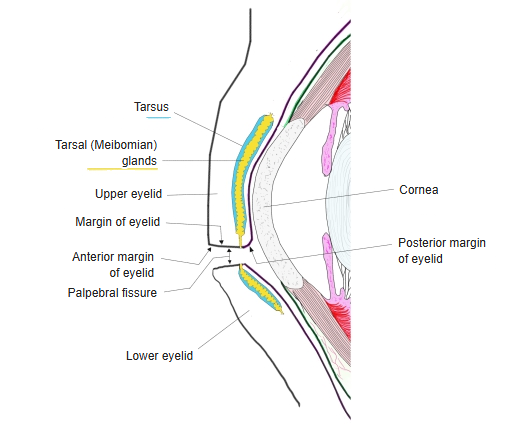
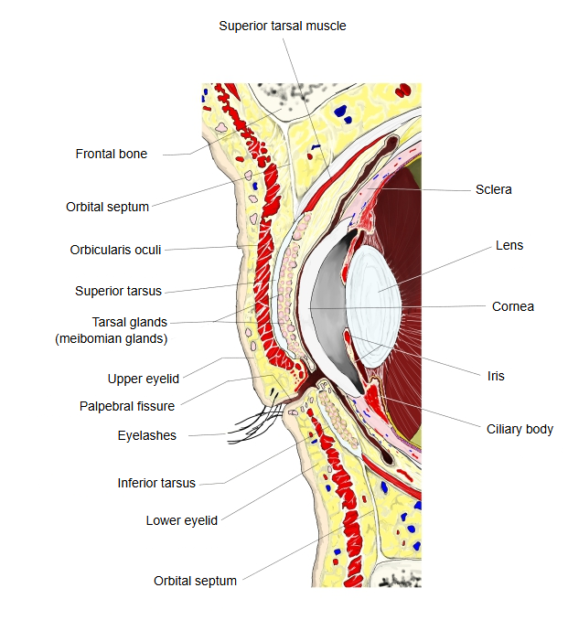
· Each gland is a straight tube with many small side pouches, set vertically in the tarsus. There are more and longer glands in the upper lid than in the lower lid; sources differ on the numbers (roughly 25 to 40 above and 20 to 30 below).
· Their ducts open on the edge of the eyelid, just in front of its posterior margin. They are not connected to the eyelash follicles.
· They produce an oily secretion (meibum) by holocrine secretion. Blinking spreads it as the outer lipid layer of the tear film, which slows evaporation, keeps tears from spilling over the lid margin and seals the closed lids.
· Poor function of these glands is a common cause of dry eye; a blocked gland can swell into a chalazion.
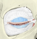 Tarsal glands (left eyelids)
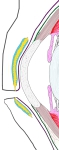 Eyelids (diagram)
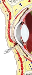 Eyelids (sagittal section)
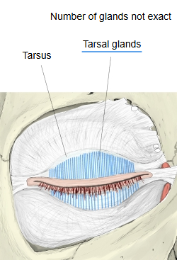
Related terms
- Edge of eyelid
- Eyelids
- Upper eyelid
- Lower eyelid
- Inferior tarsus
- Holocrine secretion
- Sebaceous glands
References and sources
- Further reading: Meibomian gland, Wikipedia.
- Federative International Programme for Anatomical Terminology (FIPAT). Terminologia Anatomica, 2nd ed. (TA2), 2019.