Tectorial membrane of cochlear duct (Latin: membrana tectoria ductus cochlearis)
Also called the tectorial membrane (cochlea)
· A membrane of the same name exists in the neck: the tectorial membrane of the craniovertebral joints.
Reference websites
[Wikipedia]
· Tectorial membrane, Wikipedia.
· Like the basilar membrane, it is a membrane that contains no cells.
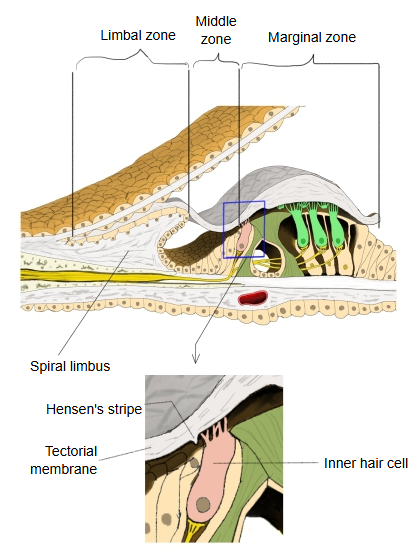
· It has three zones (see the figure below):
1. 1. Limbal zone: the thinnest part, lying on the upper surface of the spiral limbus.
2. 2. Middle zone: the thickest part, over the internal spiral sulcus and the inner hair cells.
3. 3. Marginal zone: the outer part, over the outer hair cells; it thins toward its free edge. Hensen's stripe runs on the underside of the middle zone, above the inner hair cells.
· It is a gel that is about 97% water.
Its dry weight is about 50% collagen, 25% non-collagenous glycoproteins (α-tectorin, β-tectorin and otogelin) and 25% proteoglycans.
Structure
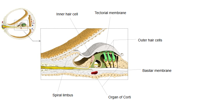
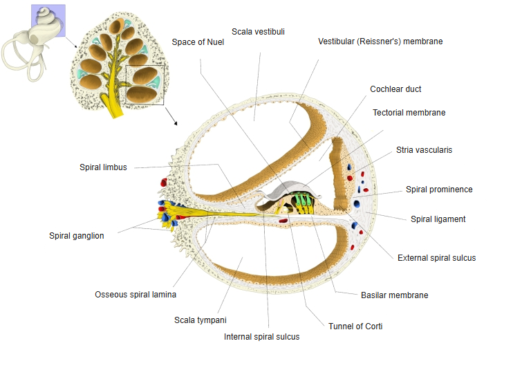
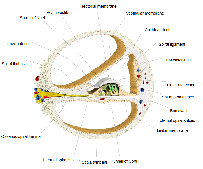
The collagen fibres of the tectorial membrane are held in a regular striated sheet matrix formed by α- and β-tectorin, proteins found only in the inner ear. Because of this structure its mechanical properties differ with direction (radially and along the cochlea) and it behaves as a viscoelastic material.
Function
Its exact mechanical role in hearing is still being studied. The tallest stereocilia of the outer hair cells are attached to it, while the inner hair cells are stimulated through the fluid between it and the organ of Corti. Studies in mice lacking tectorin proteins show changes in hearing, and isolated tectorial membrane can carry travelling waves at sound frequencies. It also stores calcium ions, which the sensory cells need to respond normally.
Related terms
- Organ of Corti
- Basilar membrane
- Spiral limbus
- Inner hair cell
- Outer hair cell
- Internal spiral sulcus
- Cochlear duct
References and sources
- Further reading: Tectorial membrane, Wikipedia.
- Federative International Programme for Anatomical Terminology (FIPAT). Terminologia Anatomica, 2nd ed. (TA2), 2019.