Basilar membrane (Latin: lamina basilaris)
Also called the basilar lamina
- Spiral canal of the cochlea : total length about 3 cm, about 2.5–2.75 turns.
- Upper level
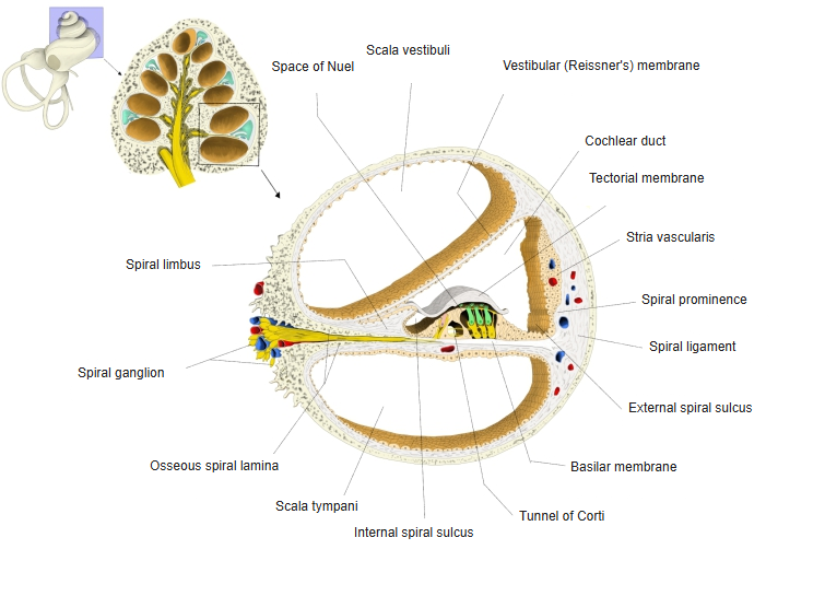
- Scala vestibuli : filled with perilymph; carries the sound vibrations to the cochlear duct; part of the bony labyrinth. Joined to the scala tympani at the apex through the helicotrema.
- Cochlear duct : separated from the scala vestibuli by the vestibular (Reissner's) membrane; filled with endolymph and contains the spiral organ (organ of Corti); both ends are blind. Contents: tectorial membrane, organ of Corti, internal and external spiral sulci, spiral prominence, stria vascularis.
- Lower level
- Scala tympani : part of the bony labyrinth, filled with perilymph; passes the travelling wave on to the tympanic cavity. Its boundary with the upper level: the osseous spiral lamina + the basilar membrane (+ the secondary spiral lamina).
- Upper level
- Modiolus : the cone of spongy bone at the centre of the cochlea, with the longitudinal canals and the spiral canal of the modiolus.
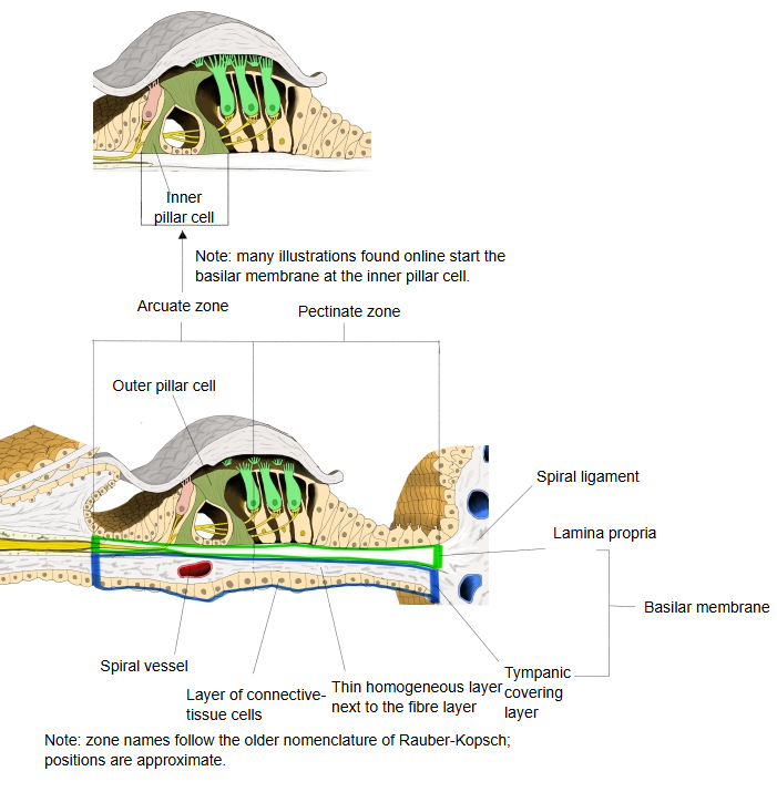
| 1 | Lamina propria | Arcuate zone | The inner part, next to the tympanic lip (the tip of the osseous spiral lamina). From the tympanic lip to the foot of the outer pillar cells; thin, with fine radial strands. |
| Pectinate zone | The outer part, from the outer pillar cells to the spiral ligament. Apart from its epithelium it has three layers: a central homogeneous layer between two fibre layers. The lower fibre layer has thicker fibres, round in cross-section. At the spiral ligament the homogeneous layer ends and only the fibre layers continue into the ligament. A few elongated nuclei are scattered in the fibre layers. | ||
| 2 | Scala tympani covering layer | Two layers: 1. a thin homogeneous layer next to the lower fibre layer; 2. a few layers of connective tissue cells rich in cytoplasm. A little lateral to the tympanic lip runs a small vessel, the spiral vessel, along the whole length of the cochlear duct. | |
Note: some textbooks divide the membrane into the arcuate and pectinate zones by the position of the organ of Corti, which differs considerably from the division above. Many modern drawings start the membrane at the inner pillar cell; sources differ in how they name and divide these parts.
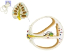 Section of a cochlear turn
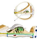 Structure of the basilar membrane
Reference websites
[Wikipedia]
· Basilar membrane, Wikipedia.
· Cochlea, Wikipedia.
[Websites with illustrations]
· Cross-section of the cochlea (Wikimedia Commons)

· Longitudinal section of the cochlea (Gray's Anatomy, fig. 928) (Wikimedia Commons)

· The inner ear (TeachMeAnatomy)
Structure
The basilar membrane behaves somewhat like the strings of an instrument, which differ in width and stiffness. Unlike the separate strings of a guitar, however, it is a single structure whose width, stiffness, mass and damping change gradually along its length.
Sound sets up a travelling wave along the membrane. The properties of each point decide its characteristic frequency, the frequency to which that point responds most strongly.
The membrane is widest (0.42–0.65 mm) and least stiff at the apex of the cochlea, and narrowest (0.08–0.16 mm) and stiffest at the base, near the oval and round windows. High-pitched sounds therefore move it most near the base, and low-pitched sounds near the apex.
Function
Separation of endolymph and perilymph: together with the vestibular membrane, the tissues carried by the basilar membrane (the cells of the inner and outer sulci and the reticular lamina of the organ of Corti) keep the endolymph of the cochlear duct apart from the perilymph. The basilar membrane itself lets perilymph through under the spiral organ, so there the border between the two fluids is the reticular lamina on the endolymph side of the organ.
Related terms
- Cochlear duct
- Spiral organ (organ of Corti)
- Cochlea
- External spiral sulcus
- Spiral canal of modiolus
- Base of cochlea
- Cochlear cupula
References and sources
- Further reading: Basilar membrane, Wikipedia.
- Further reading: Cochlea, Wikipedia.
- Federative International Programme for Anatomical Terminology (FIPAT). Terminologia Anatomica, 2nd ed. (TA2), 2019.