Third ventricle (Latin: ventriculus tertius)
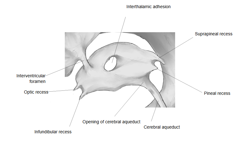
What is the third ventricle?
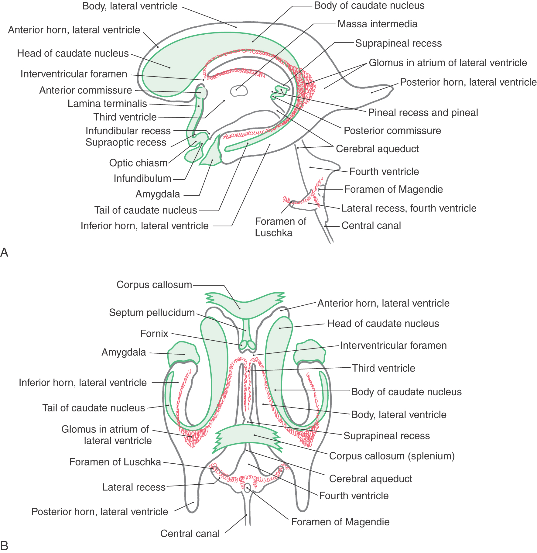
The brain has four ventricles in all: the two lateral ventricles (one in each cerebral hemisphere), the third ventricle and the fourth ventricle.
⇒ An illustration showing the flattened shape (Wikimedia Commons)
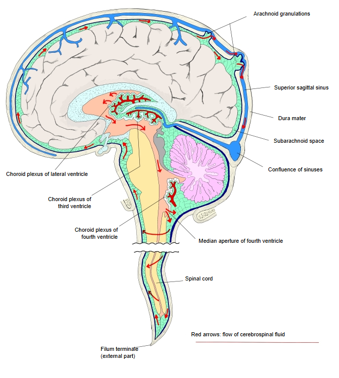
The ventricles are not separate from each other: they are connected, and they produce and secrete cerebrospinal fluid and pass it on. The fluid flows from the lateral ventricles into the third ventricle, then through the cerebral aqueduct to the fourth ventricle, and leaves through its median and lateral apertures into the subarachnoid space.
The choroid plexus of the third ventricle forms two thin strips that run from front to back along the roof of the ventricle; at each side it is attached along the stria medullaris thalami by the taenia thalami. At the interventricular foramina it is continuous with the choroid plexus of the lateral ventricles.
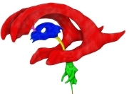 Ventricles: left side (coloured)
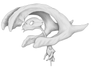 Ventricles: left side
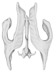 Ventricles: from above
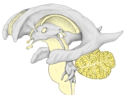 Third ventricle and surroundings
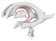 Choroid plexuses
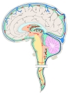 Flow of cerebrospinal fluid
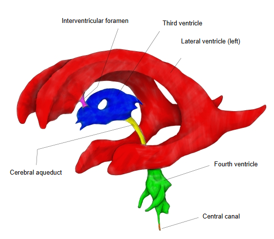
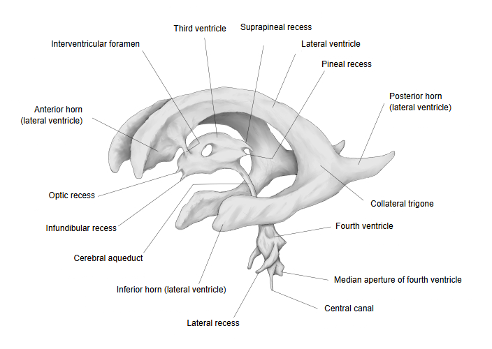
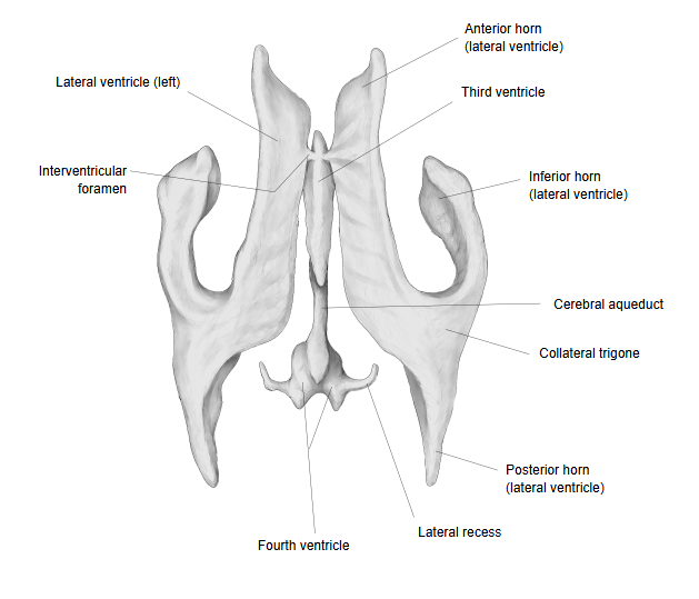
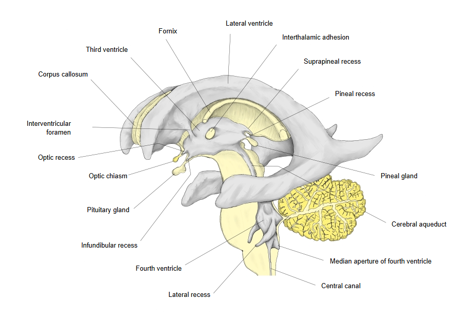
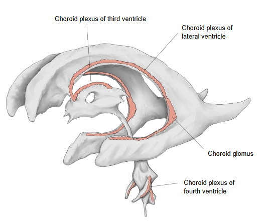

Walls of the third ventricle
The walls of the third ventricle are as follows.
Roof: formed by the tela choroidea of the third ventricle with the choroid plexus of the third ventricle and the thin ependymal (epithelial) layer that covers its underside. At each side the roof is attached along the stria medullaris thalami by the taenia thalami; behind, it attaches to the habenulae and the upper surface of the pineal gland, so the ventricle forms a pocket above the pineal gland: the suprapineal recess.
Posterior wall (upper part): formed by the stalk of the pineal gland and the posterior commissure; the ventricle runs back into the pineal stalk as the pineal recess.
Just below the pineal gland lies the posterior commissure, a bundle of white-matter fibres that links structures of the midbrain and diencephalon (for example the pretectal areas); it is not a commissure between the two cerebral hemispheres. Just below it the ventricle opens into the cerebral aqueduct.
Floor (inferior wall): formed mainly by the hypothalamus (from front to back the optic chiasm, the infundibulum, the tuber cinereum and the mammillary bodies); it forms the infundibular recess, down into the stalk of the pituitary gland, and the optic recess, in front of and above the optic chiasm.
Anterior wall: formed by the columns of the fornix, the anterior commissure and the lamina terminalis.
Hypothalamic sulcus
· Hypothalamic sulcus: a shallow groove on the lateral wall of the third ventricle that runs from the interventricular foramen to the opening of the cerebral aqueduct. It separates the medial surface of the thalamus (above) from that of the hypothalamus (below).
Reference websites
[Wikipedia]
· Third ventricle, Wikipedia.
· Ventricular system, Wikipedia.
[Websites with illustrations]
· Ventricles of the brain: lateral and top views with recesses and choroid plexus (ClinicalGate)
· Third ventricle (brain) (Kenhub)
· The ventricles of the brain (TeachMeAnatomy)
Where it is found
Related terms
References and sources
- Further reading: Third ventricle, Wikipedia.
- Further reading: Ventricular system, Wikipedia.
- Federative International Programme for Anatomical Terminology (FIPAT). Terminologia Anatomica, 2nd ed. (TA2), 2019.