Cerebral ventricles (Latin: ventriculi cerebri)
Also called the ventricles of the brain or the ventricular system
· Overview
Overview
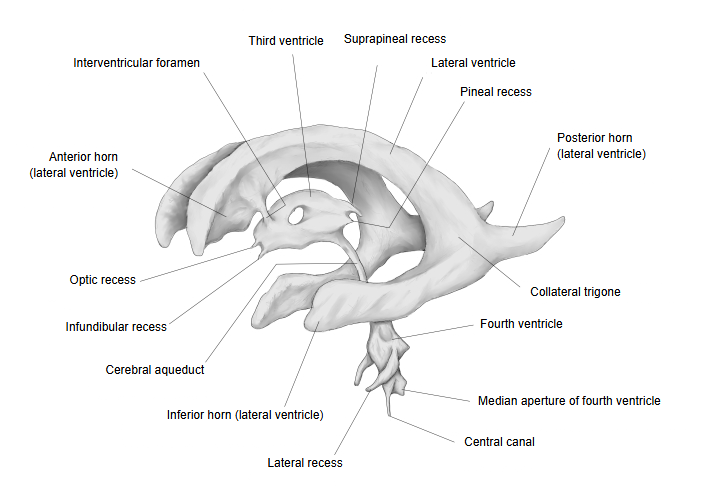
The cerebral ventricles are
ventricles
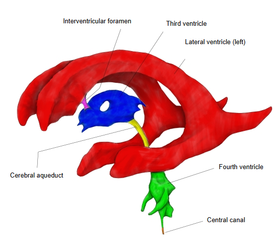
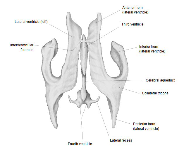
- Lateral ventricles : one in each cerebral hemisphere, right and left: the left and right lateral ventricles.
- Third ventricle : in the midline of the diencephalon.
- Fourth ventricle : between the medulla oblongata and pons in front and the cerebellum behind.
· The ventricles are lined by ependyma and continue below into the central canal of the spinal cord. They develop from the central cavity of the embryonic neural tube.
· CSF is made mainly by the choroid plexus in each ventricle. It flows from the lateral ventricles to the third ventricle, through the cerebral aqueduct to the fourth ventricle, and out into the subarachnoid space, where it is absorbed into the venous sinuses.
Lateral ventricles
· One in each cerebral hemisphere; the largest ventricles, C-shaped.
· Parts: anterior horn (frontal lobe), central part (parietal lobe), posterior horn (occipital lobe) and inferior horn (temporal lobe). The central part, posterior horn and inferior horn meet at the collateral trigone (atrium).
· Each opens into the third ventricle through the interventricular foramen.
Third ventricle
· A narrow slit in the midline of the diencephalon, between the right and left thalami and hypothalamus. It has small recesses: the optic, infundibular, pineal and suprapineal recesses.
· Behind and below, it continues into the cerebral aqueduct, a narrow canal through the midbrain. See Third ventricle.
Sources differ on whether the third ventricle is described as part of the brainstem: it lies in the diencephalon, which some texts include in the brainstem and others do not.
Fourth ventricle
· A tent-shaped cavity between the pons and medulla oblongata in front and the cerebellum behind. Its floor is the rhomboid fossa; see also roof of the fourth ventricle.
· It opens into the subarachnoid space through the median aperture and the two lateral apertures, at the ends of the lateral recesses. Below, it narrows into the central canal of the medulla and spinal cord.
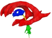 Ventricles: left side (coloured)
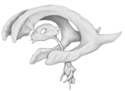 Ventricles: left side
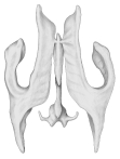 Ventricles: from above
Useful sites
· Ventricles of the brain (Blausen, Wikimedia Commons)

· Circulation of cerebrospinal fluid (Wikimedia Commons)

· Ventricular system (Wikipedia)
Related terms
- Pia mater
- Rhomboid fossa
- Third ventricle
- Inferior horn of lateral ventricle
- Posterior horn of lateral ventricle
- Choroid plexus
- Subarachnoid space
- Brain
References and sources
- Further reading: Ventricular system, Wikipedia.
- Federative International Programme for Anatomical Terminology (FIPAT). Terminologia Anatomica, 2nd ed. (TA2), 2019.