Median atlanto-axial joint (Latin: articulatio atlantoaxialis mediana)
What is the median atlanto-axial joint?
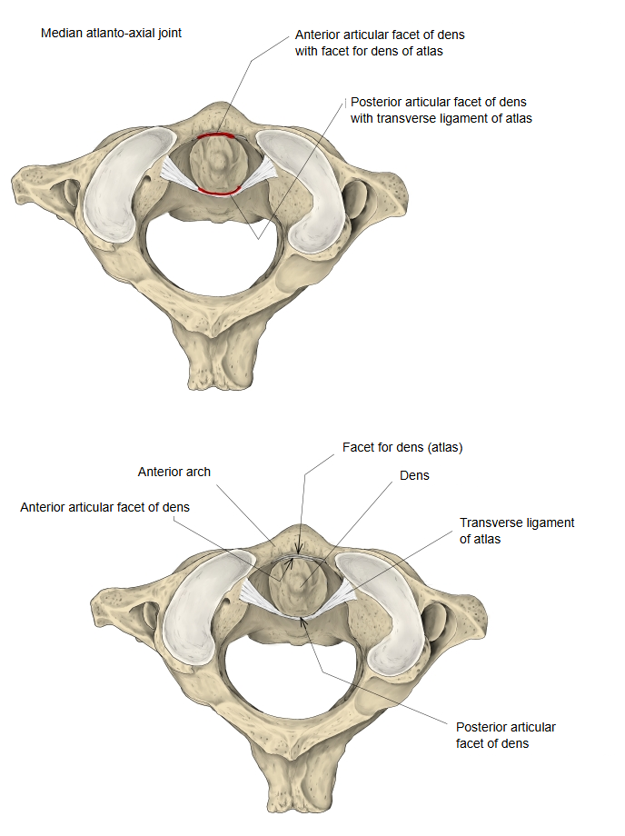
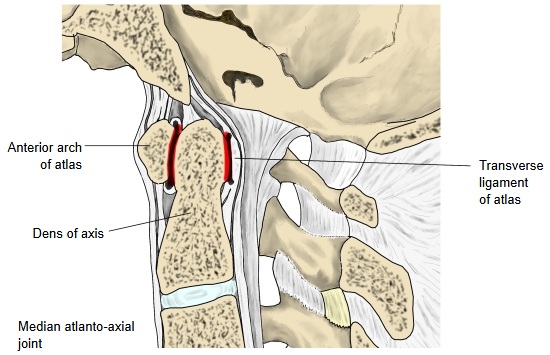
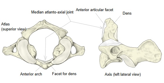
Front part: atlas (anterior arch, facet for dens) ⇔ axis (dens, anterior articular facet)
Back part: axis (dens, posterior articular facet) ⇔ transverse ligament of atlas
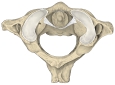 Superior view
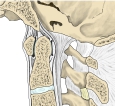 Median section
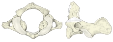 Articular surfaces
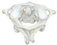 Atlas + axis
 Related ligaments
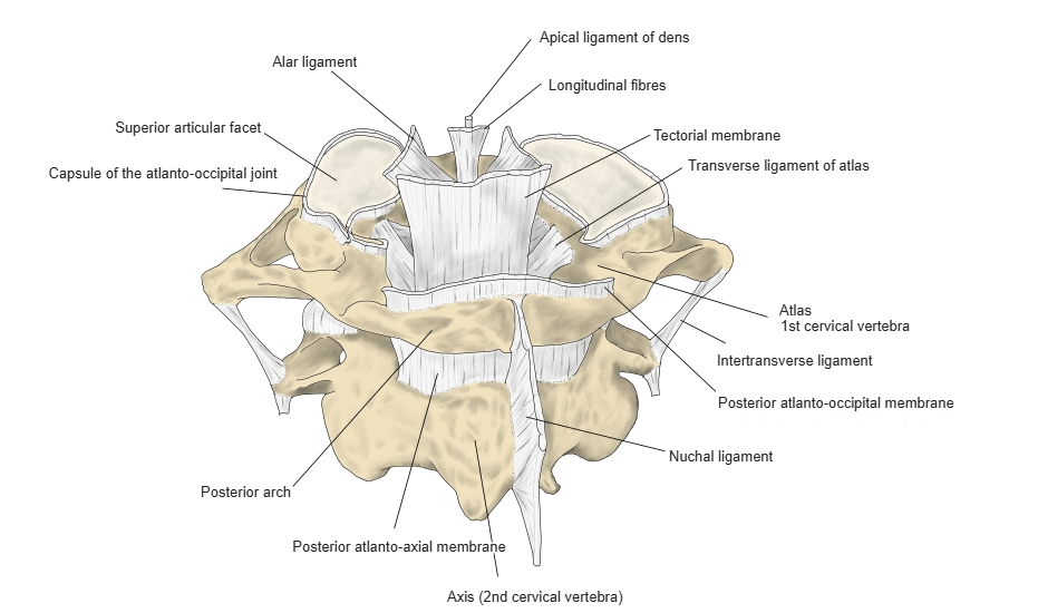
- Each joint capsule is thin and loose.
- The surface of the transverse ligament of atlas that faces the back joint cavity is covered with fibrocartilage.
- Together with the lateral atlanto-axial joints, it lets the atlas, carrying the skull, rotate (turning the head to the side).
Bones
The joint has two parts:
Back part: axis (dens, posterior articular facet) ⇔ transverse ligament of atlas
More about the joint
The median atlanto-axial joint is a pivot joint made up of two small, separate joints: one between the anterior articular facet of the dens and the facet for dens on the back of the anterior arch of the atlas, and one between the posterior articular facet of the dens and the transverse ligament of atlas. Each has a thin, loose capsule, and the surface of the transverse ligament facing the back cavity is covered with fibrocartilage. Because this joint, together with the lateral atlanto-axial joints, rotates the atlas and the skull on the axis, its build is unlike that of other joints: the dens fits like an axle into a ring formed by the back of the anterior arch of the atlas and the transverse ligament, and the atlas turns around the dens.
The following ligaments control this pivot joint:
- Cruciform ligament of atlas: a cross-shaped ligament covering the back of the dens. It has two parts. (a) The transverse ligament of atlas stretches between the right and left lateral masses of the atlas; it is an articular surface and at the same time stops the dens from slipping backwards. It is unusual in running between two points on the same bone. (b) The longitudinal bands run up and down from the middle of the transverse ligament, to the anterior margin of the foramen magnum and to the back of the body of the axis.
- Alar ligaments: arise from both sides of the upper part of the dens and run upwards and outwards to the medial sides of the occipital condyles. They have no attachment to the atlas. They are strong, thick, short ligaments and limit excessive rotation of the head.
- Apical ligament of dens: a thin ligament joining the tip of the dens to the anterior margin of the foramen magnum. It does little to control movement, but it is important as a remnant marking the position of the notochord.
- Tectorial membrane: a broad membrane covering the cruciform ligament from behind. It reaches the clivus and blends with the dura mater. It is regarded as the upward continuation of the posterior longitudinal ligament.