Latin: musculus abductor digiti minimi pedis
Overview
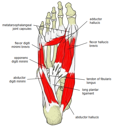
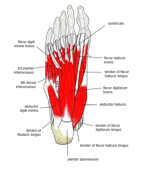
- One of the 3 muscles of the little toe in the sole (with the flexor digiti minimi brevis and opponens digiti minimi).
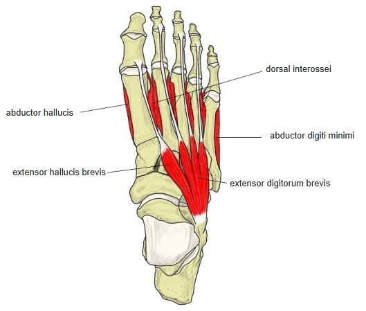
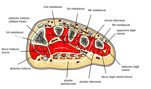
- Lies along the lateral border of the foot, just under the skin of the sole.
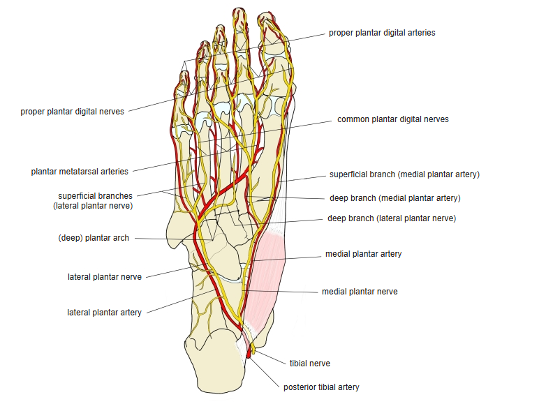
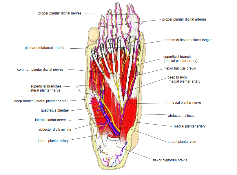
- Its medial margin is related to the lateral plantar artery, vein and nerve.
- It corresponds to the abductor digiti minimi of the hand.
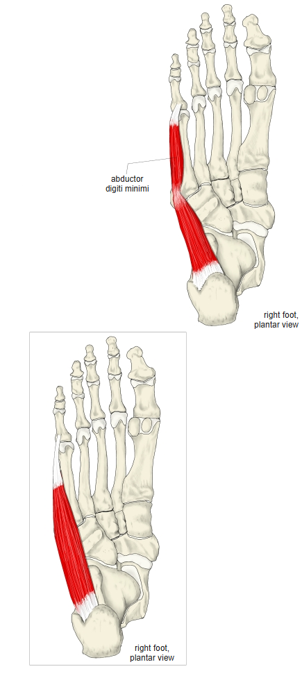
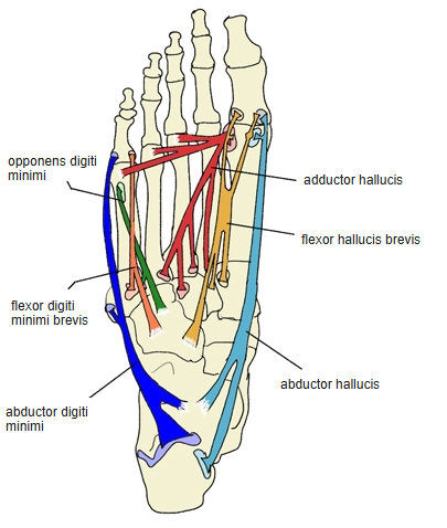
- The figure follows the Prometheus atlas, which shows the muscle with two bellies; many other illustrations show a single belly.
Position in the classification
Lower Limb › Foot › Sole of foot › Muscles of the little toe
- Abductor digiti minimi (foot)
- Flexor digiti minimi brevis (foot)
- Opponens digiti minimi (foot)
Illustrations
Origin / insertion
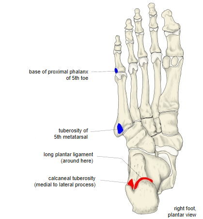
- Origin: calcaneus — the lateral and medial processes of the calcaneal tuberosity and the under surface between them — the plantar aponeurosis and the intermuscular septum next to the flexor digitorum brevis
- Some sources also list the long plantar ligament or the tuberosity of the 5th metatarsal.
- Insertion: lateral side of the base of the proximal phalanx of the 5th toe, together with the flexor digiti minimi brevis
- Some sources also give the tuberosity of the 5th metatarsal as an insertion; others describe the tendon gliding over it.
Action
- Flexes and abducts the little toe (moves it away from the 4th toe) at the metatarsophalangeal joint
- When the foot bears weight, helps to support the lateral longitudinal arch
Innervation
- Lateral plantar nerve (S1–S3), a branch of the tibial nerve
Blood supply
- Lateral plantar artery
Related terms
- Muscles of the little toe
- Flexor digiti minimi brevis (foot)
- Opponens digiti minimi (foot)
- Abductor digiti minimi (hand)
- Calcaneal tuberosity
- Plantar aponeurosis
- Long plantar ligament
- Lateral plantar nerve
- Lateral plantar artery
- Tibial nerve
References and sources
- Further reading: Abductor digiti minimi muscle of foot, Wikipedia.