Latin: nervus plantaris lateralis
Overview
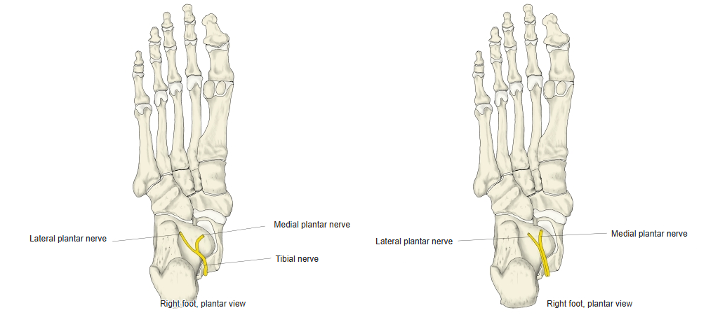
- The smaller terminal branch of the tibial nerve; comparable to the ulnar nerve in the hand.
- Supplies most of the deep muscles of the sole and the skin of the lateral sole and the lateral 1½ toes.
Division of the tibial nerve 
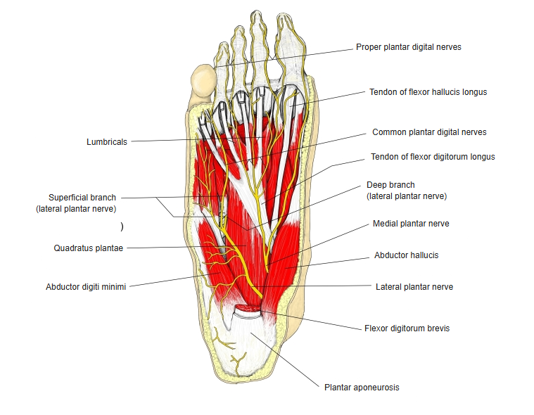
Nerves of the sole
Course
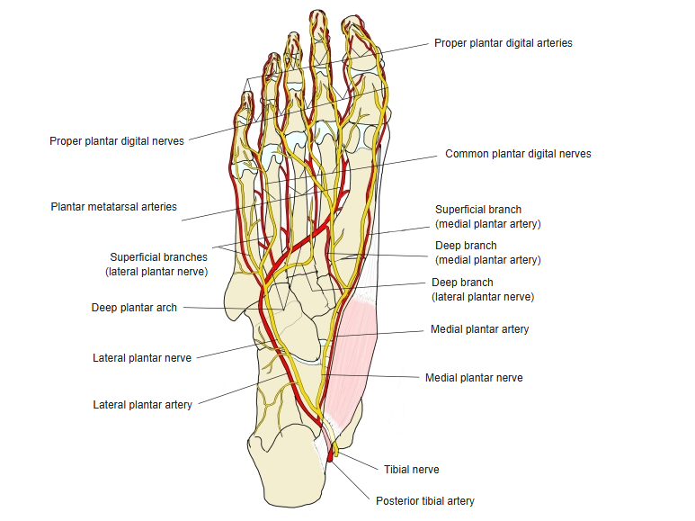
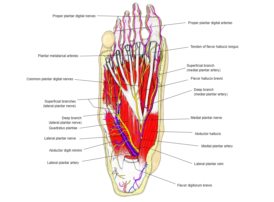
- Runs towards the little toe along the medial side of the lateral plantar artery and vein.
- Passes between flexor digitorum brevis and quadratus plantae, then between flexor digitorum brevis and abductor digiti minimi.
- Divides near the base of the 5th metatarsal into a superficial and a deep branch.
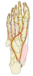
Nerves and arteries of the sole 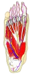
Nerves, vessels and muscles of the sole
Branches and supply
| Branch | Supplies |
|---|---|
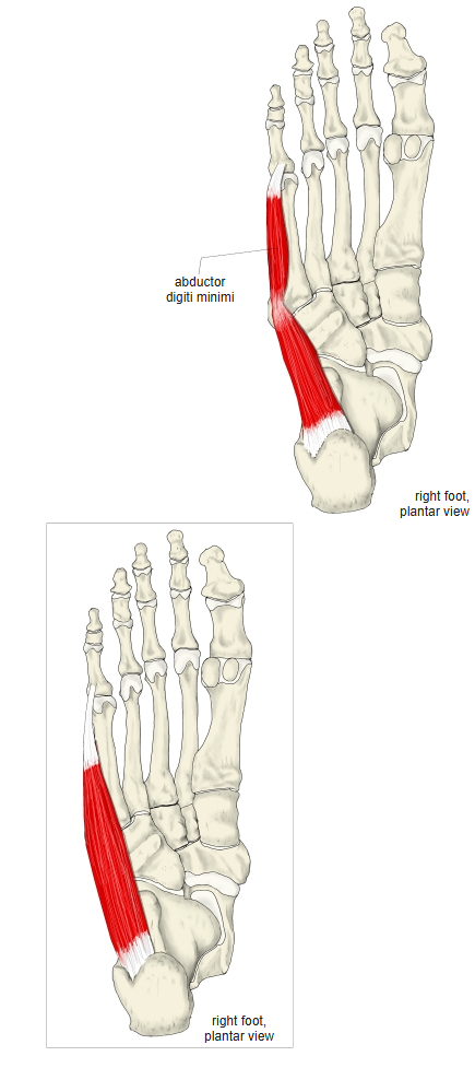
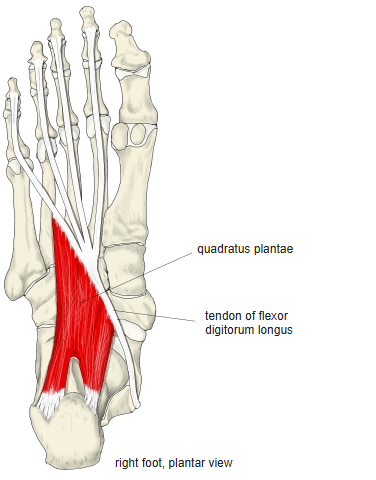
| Muscular branches (trunk) | Quadratus plantae, abductor digiti minimi |
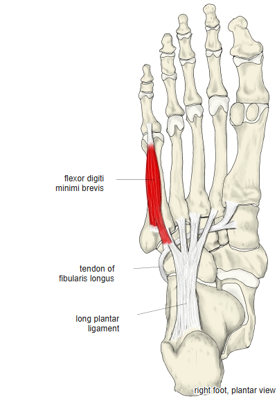
| Superficial branch | A proper plantar digital nerve to the lateral side of the little toe, and a common plantar digital nerve dividing into two proper plantar digital nerves (adjacent sides of the 4th and 5th toes); flexor digiti minimi brevis |
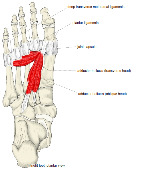
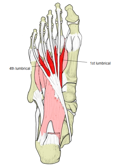
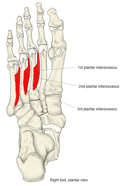
| Deep branch | Arches medially with the plantar arterial arch: adductor hallucis, the interossei, the lateral three lumbricals |
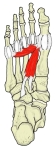
Adductor hallucis 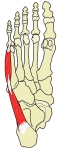
Abductor digiti minimi (foot) 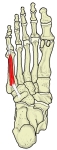
Flexor digiti minimi brevis (foot) 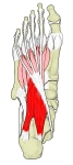
Quadratus plantae 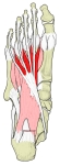
Lumbricals of the foot 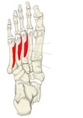
Plantar interossei
References and sources
- Further reading: Lateral plantar nerve, Wikipedia.