Latin: musculus brachialis
Overview
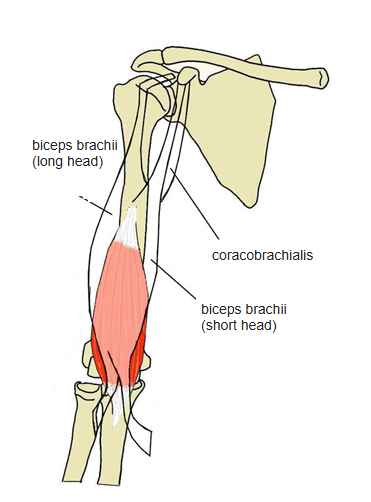
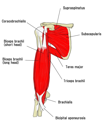
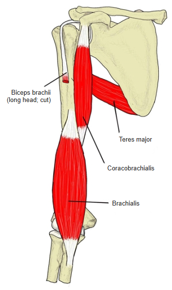
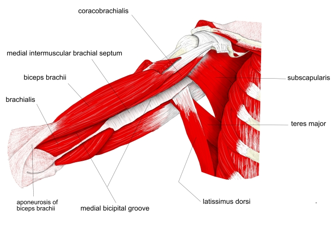
- One of the 3 muscles of the anterior (flexor) compartment of the arm (with the biceps brachii and coracobrachialis).
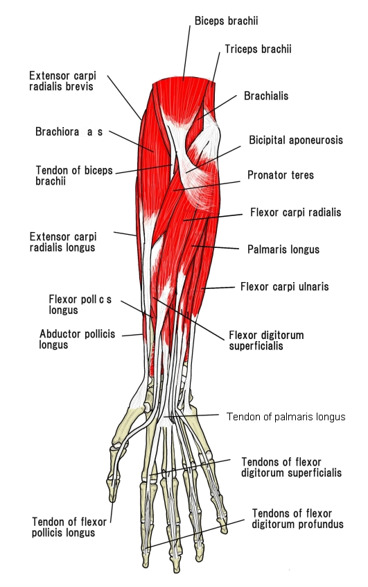
- Lies deep to the biceps brachii and forms part of the floor of the cubital fossa.
- The most powerful flexor of the elbow: it produces more force than the biceps.
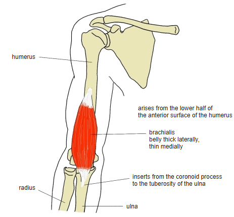
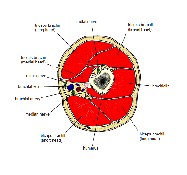
- Its belly is thick laterally and thin medially.
- Lying directly on the humerus, it is easily injured by pressure or by the bone ends in a supracondylar fracture; scarring can shorten it so that the elbow cannot be fully extended.
- Continuous in places with the triceps brachii, deltoid and coracobrachialis.
Palpation
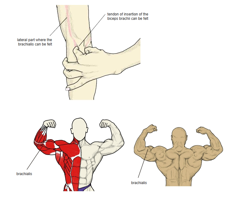
- The part not covered by the biceps can be felt under the skin.
- Pinch the biceps tendon at the elbow between thumb and index finger: the lowest part of the brachialis can be felt just beneath it. Following it upward, the lateral and medial bellies can be felt under the biceps.
Position in the classification
Upper Limb › Arm › Flexor group
- Biceps brachii
- Coracobrachialis
- Brachialis
Illustrations
Origin / insertion
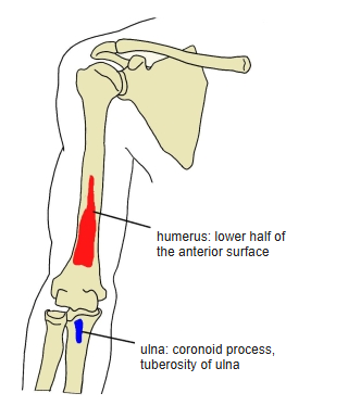
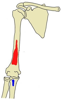
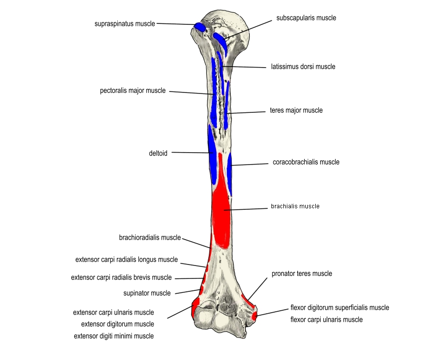
- Origin: lower half of the anterior surface of the humerus, the medial and lateral intermuscular septa, and the front of the elbow-joint capsule
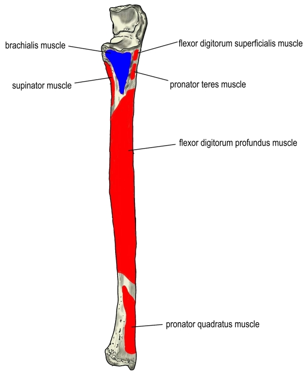
- Insertion: coronoid process and tuberosity of the ulna, and the front of the elbow-joint capsule
Action
- Flexes the elbow (the prime mover of elbow flexion)
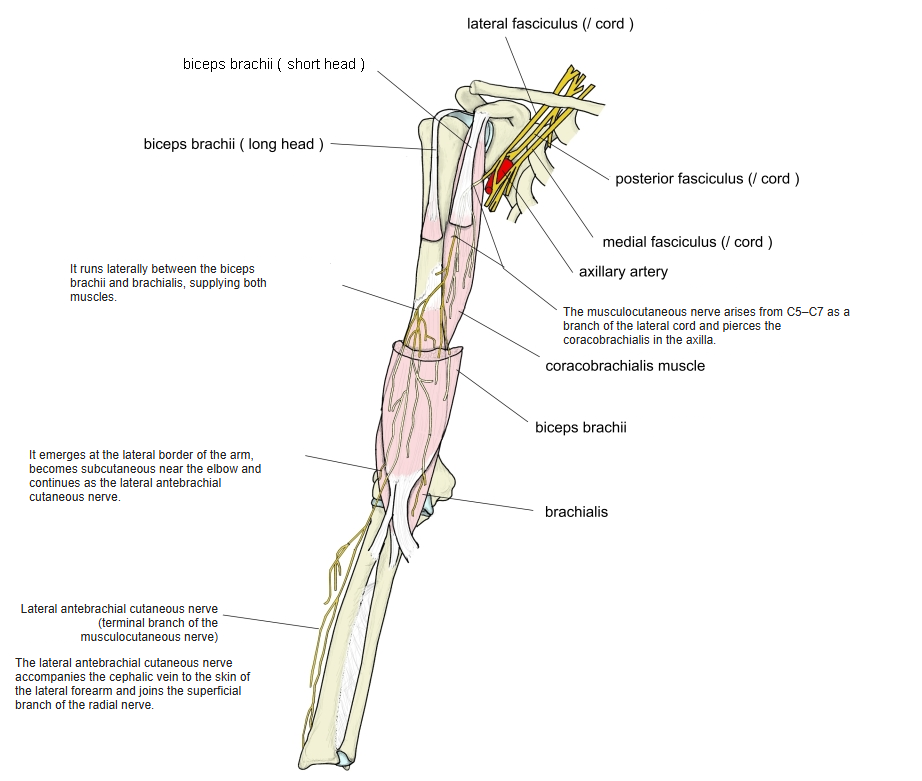
Innervation
- Musculocutaneous nerve (C5–C6)
- Part of the lateral portion: radial nerve (C7)
Blood supply
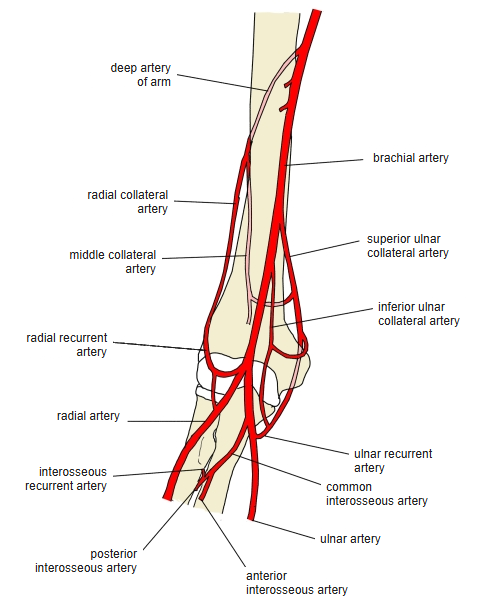
- Muscular branches of the brachial artery, the superior and inferior ulnar collateral arteries, and the radial recurrent artery
Related terms
- Biceps brachii
- Coracobrachialis
- Triceps brachii
- Humerus
- Coronoid process of ulna
- Tuberosity of ulna
- Cubital fossa
- Musculocutaneous nerve
- Radial nerve
- Brachial artery
References and sources
- Further reading: Brachialis muscle, Wikipedia.