Latin: musculus coccygeus (ischiococcygeus)
Overview
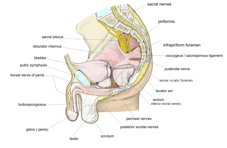
- Completes the pelvic diaphragm behind the levator ani; lies in front of the sacrospinous ligament.
- Some textbooks count it with the anal muscles, as a part of the levator ani group.
- In humans it is a reduced muscle; its superficial part is represented by the sacrospinous ligament.
- It contains so much tendon that it may look like part of the sacrospinous ligament; it may be completely absent (Rauber-Kopsch).
- The pudendal nerve runs between it and the piriformis (Wikipedia).
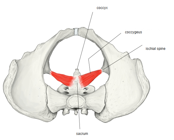
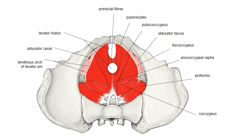
Illustrations
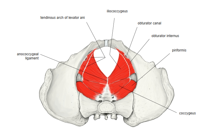
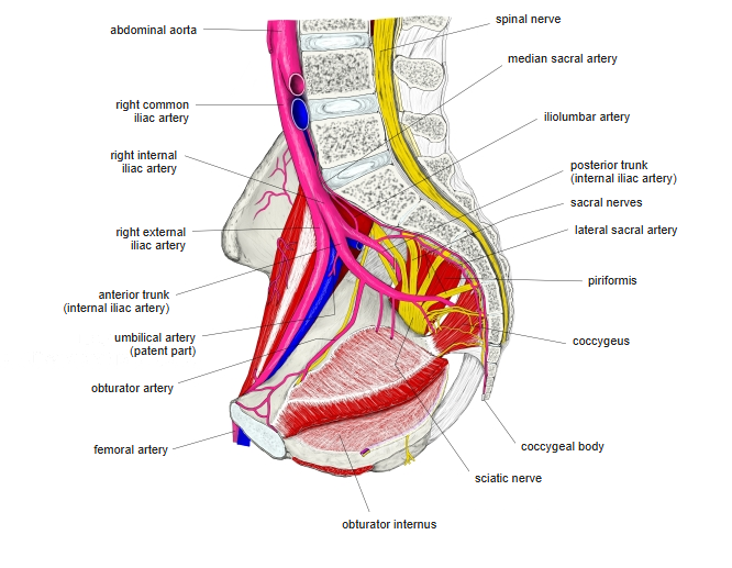
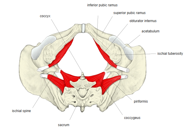
Origin / insertion
- Origin: ischial spine and the sacrospinous ligament
- Insertion: lateral margin of the coccyx and the side of the lowest part of the sacrum
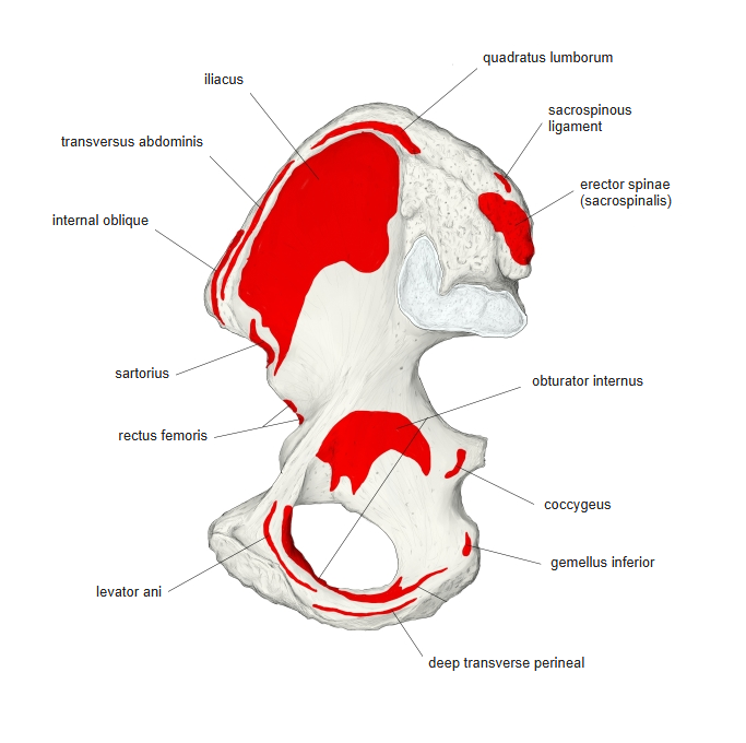
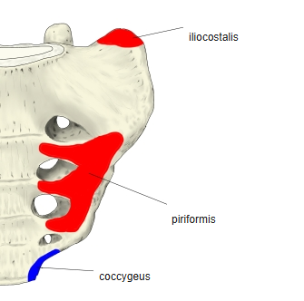
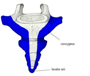
Action
- Supports the pelvic organs; with the levator ani and piriformis it closes the back part of the pelvic outlet
Innervation
- Pudendal nerve; branches of the sacral nerves (S4–S5, or S3–S4)
Blood supply
- Not given in the sources used here
Related terms
- Levator ani
- Iliococcygeus
- Piriformis
- Sacrospinous ligament
- Ischial spine
- Coccyx
- Pudendal nerve
References and sources
- Further reading: Coccygeus muscle, Wikipedia.