Latin: musculi intercostales externi
Overview
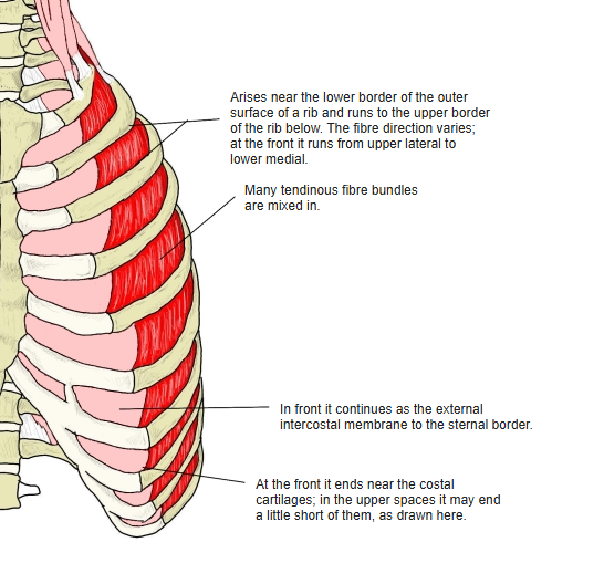
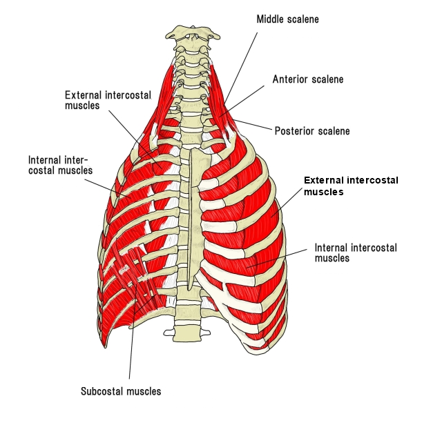
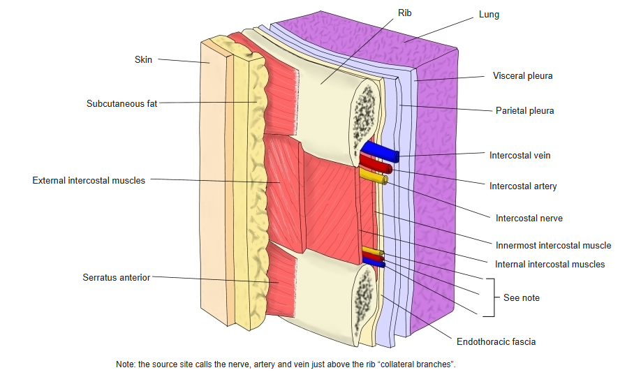
- Eleven on each side, one in each intercostal space; thicker than the internal intercostals.
- Tendinous fibres are intermingled with the muscle fibres.
- In front they end near the costal cartilages and continue as the external intercostal membrane to the sternum.
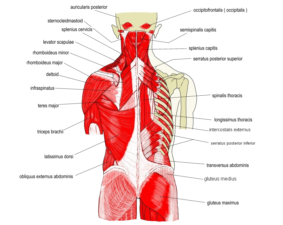
- Fibre direction: obliquely downward and lateral on the back of the thorax; downward, forward and medial on the front.
- Muscles of inspiration.
Position in the classification
Trunk › Thorax › Deep muscles of the thorax
- External intercostal muscles
- Internal intercostal muscle
- Subcostal muscle
- Levatores costarum longi
- Levatores costarum breves
- Transversus thoracis
Illustrations
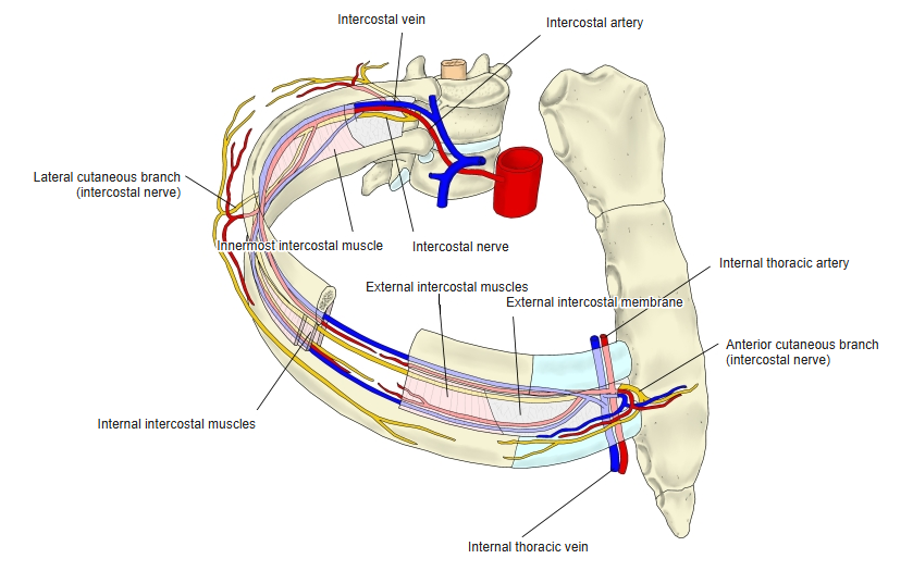
Origin / insertion
- Origin: lower border of each of ribs 1–11, from the tubercle of the rib behind to the costal cartilage in front
- Insertion: upper border of the rib below
Action
- Raise the ribs and enlarge the thoracic cavity (inspiration)
Innervation
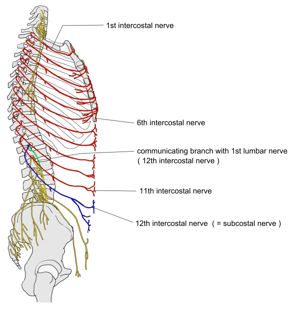
- Intercostal nerves (T1–T11)
Blood supply
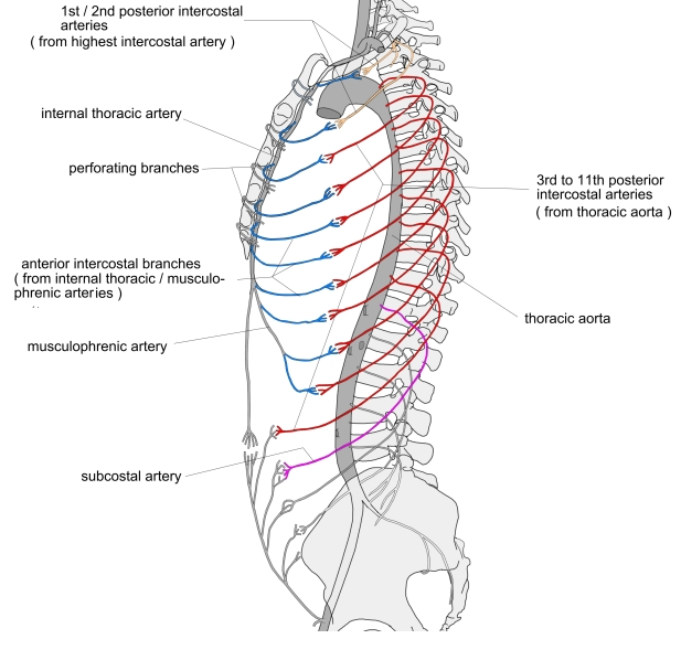
- Intercostal arteries (posterior and anterior)
Related terms
- Internal intercostal muscles
- Innermost intercostal muscles
- Diaphragm
- Intercostal nerve
- External intercostal membrane
References and sources
- Further reading: External intercostal muscles, Wikipedia.