Latin: musculi subcostales
Overview
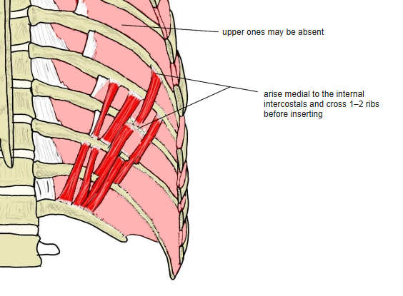
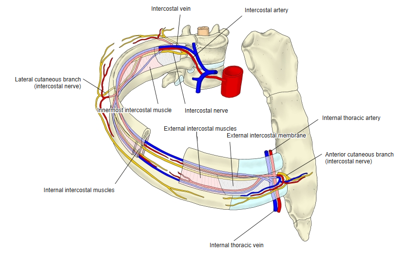
- Muscular and aponeurotic bundles on the inside of the chest wall, deep to the internal intercostals and running in the same direction.
- Best developed in the lower part of the thorax; the upper ones may be absent.
- They may be missing altogether, or join into one long band reaching from the 3rd to the 12th rib (Rauber-Kopsch).
- Some sources (e.g. Wikipedia) do not call them respiratory muscles.
Position in the classification
Trunk › Thorax › Deep muscles of the thorax
- External intercostal muscles
- Internal intercostal muscle
- Subcostal muscle
- Levatores costarum longi
- Levatores costarum breves
- Transversus thoracis
Illustrations
Origin / insertion
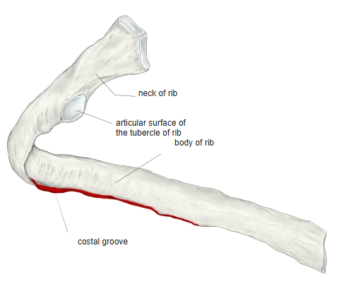
- Origin: inner surface of a rib at the upper edge of the costal groove, medial to the internal intercostals
- Insertion: the next rib below, or a rib one or two further down
Action
- Assists in lowering the ribs
Innervation
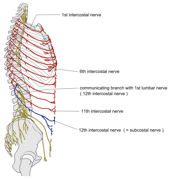
- Muscular branches of the intercostal nerves
Blood supply
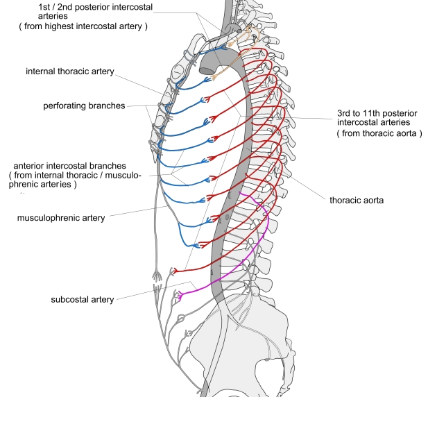
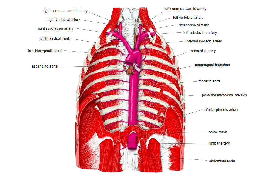
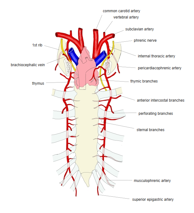
- Musculophrenic artery, posterior intercostal arteries
Related terms
- Internal intercostal muscles
- External intercostal muscles
- Transversus thoracis
- Costal groove
- Intercostal nerves
References and sources
- Further reading: Subcostales, Wikipedia.